Go back
Large lesion brain imaging with synthetic MRI
Patient information
Patient with a large brain lesion. AI based SmartSpeed is utlized to shorten scan time without compromise in image quality. Advanced imaging techniques like pCASL and 3D APT are used to perform contrast-free brain imaging to assess perfusion and tumoral activity. SWIp 3D susceptibility weighted offers the high sensitivity required to visualize deoxygenated (venous) blood or calcium deposits. A single synthetic (SyntAc) brain quantification scan is added. The resulting data of this scan can be used as input for advanced third party processing software* to synthesize MR images with different contrasts, brain parenchyma fraction maps and/or brain segmentation maps.Gallery
-
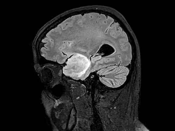
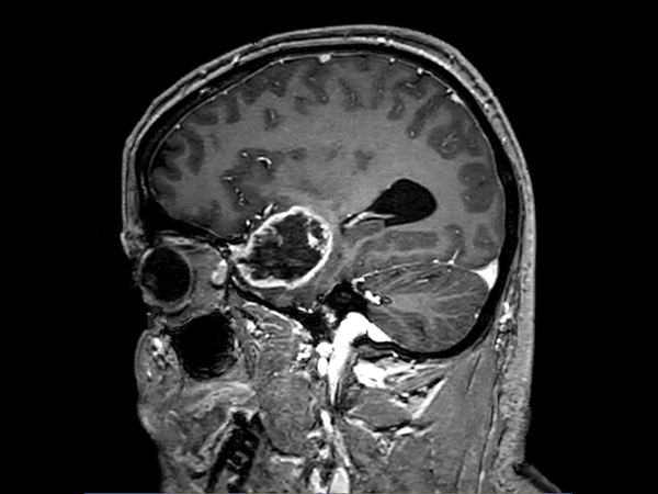
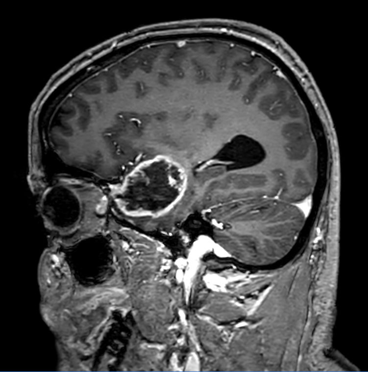
Sagittal 3D FLAIR
![Sagittal 3D FLAIR]()
-
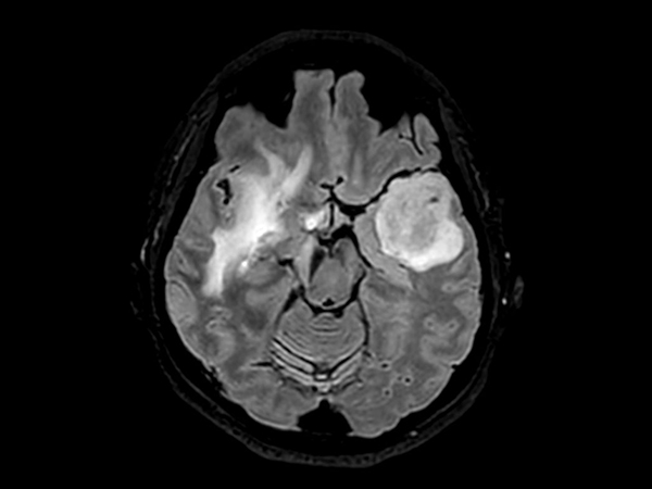
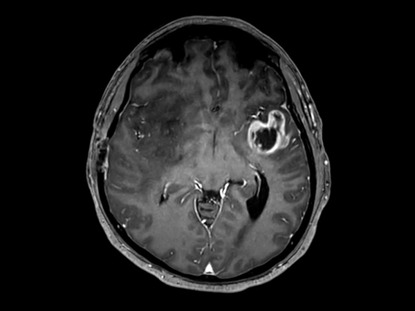
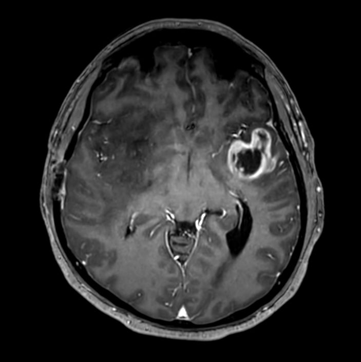
3D FLAIR (Axial reformat)
![3D FLAIR (Axial reformat)]()
-
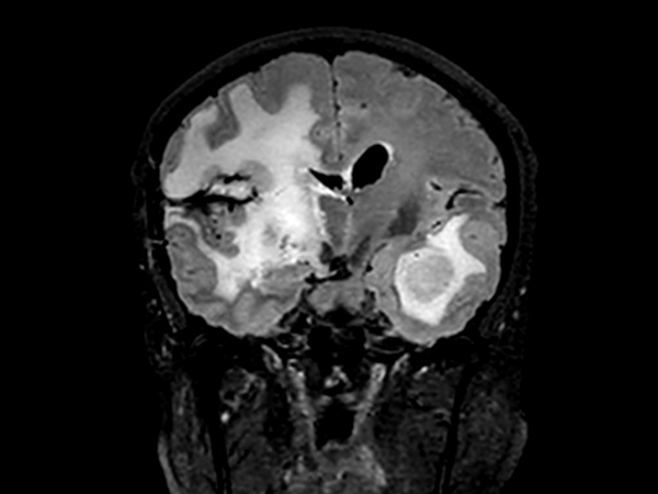
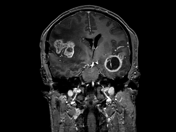
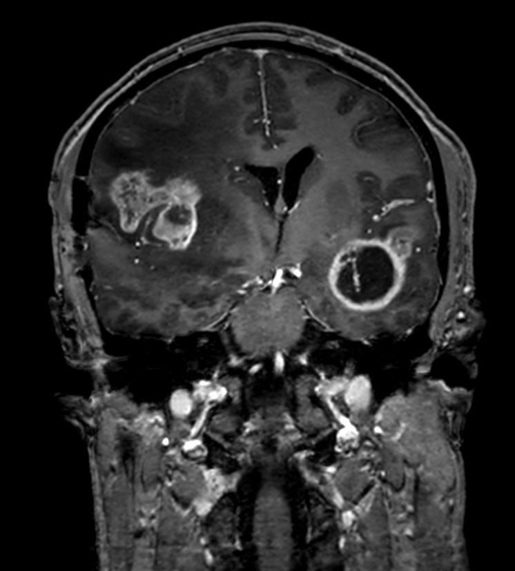
3D FLAIR (Coronal reformat)
![3D FLAIR (Coronal reformat)]()
-
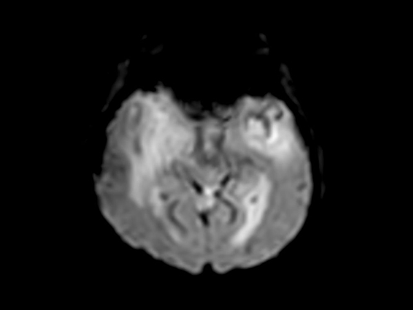
Axial T2w TSE Motion-Free
![Axial T2w TSE Motion-Free]()
-
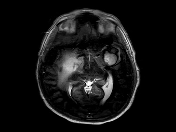
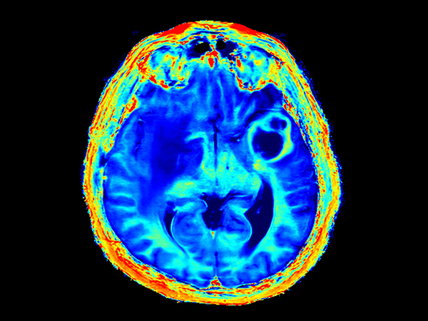
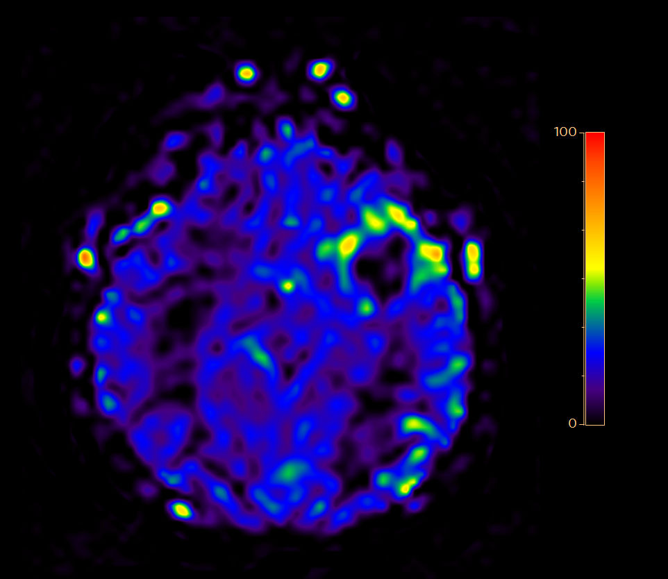
Axial 3D pCASL (3 dynamics)
![Axial 3D pCASL (3 dynamics)]()
-
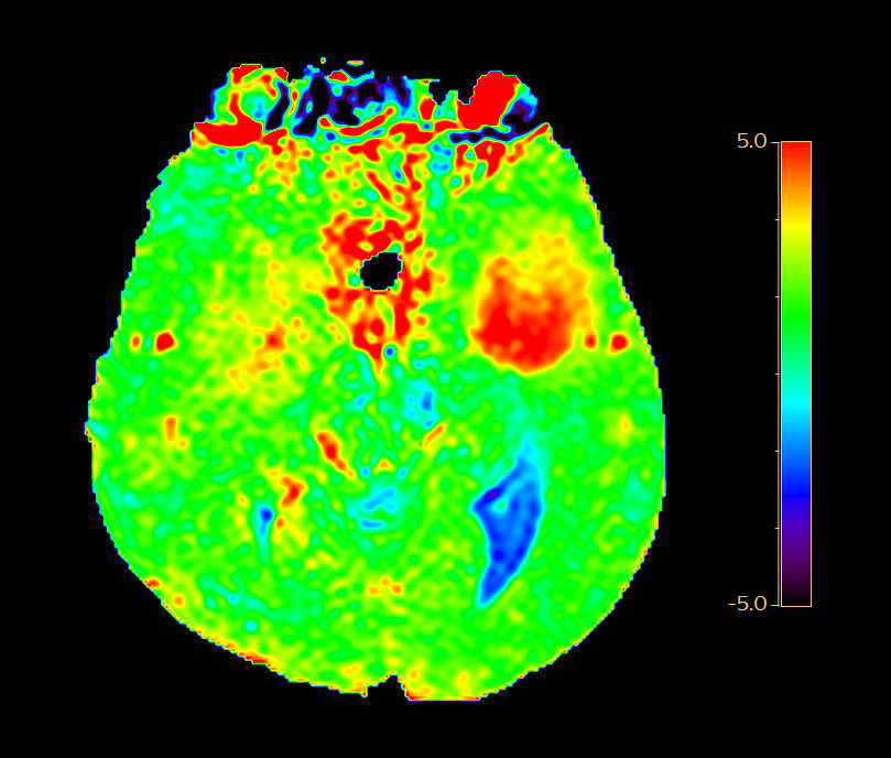
Axial 3D APT
![Axial 3D APT]()
-
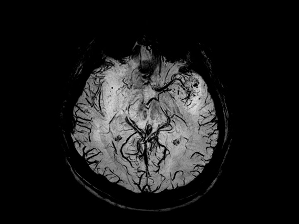
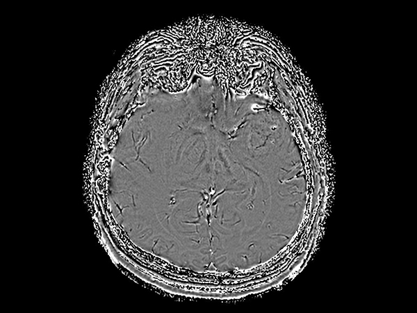
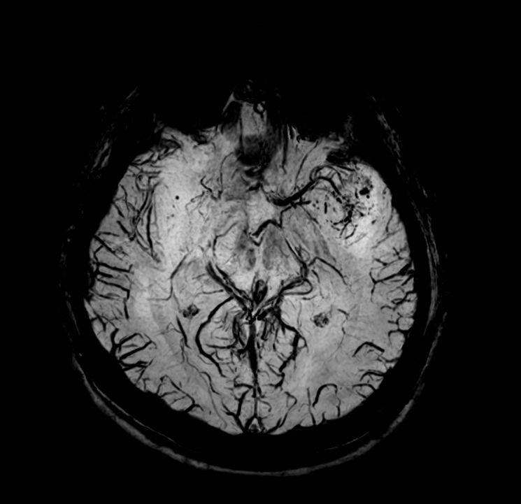
Axial SWIp (Modulus)
![Axial SWIp (Modulus)]()
-
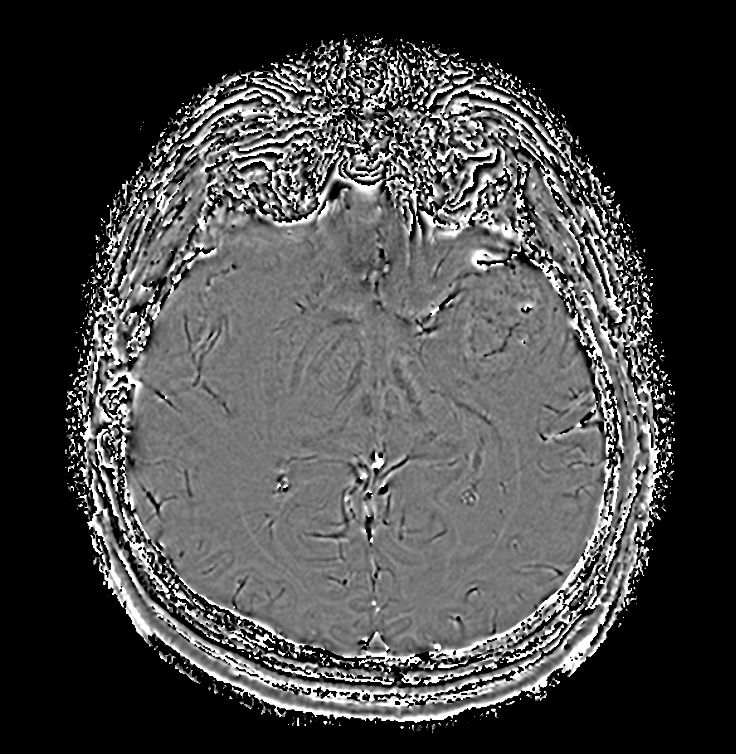
Axial SWIp (minIP)
![Axial SWIp (minIP)]()
-
Axial SWIp (Phase)
![Axial SWIp (Phase)]()
-
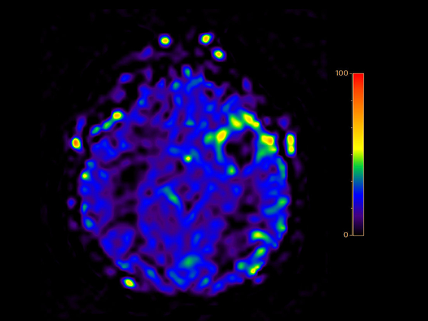
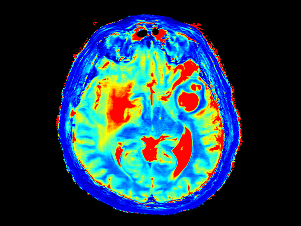
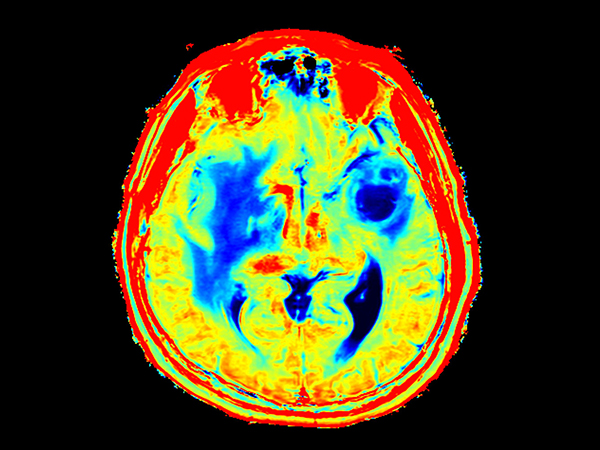
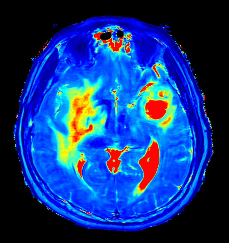
Axial T2* Perfusion (60 dynamics)
-
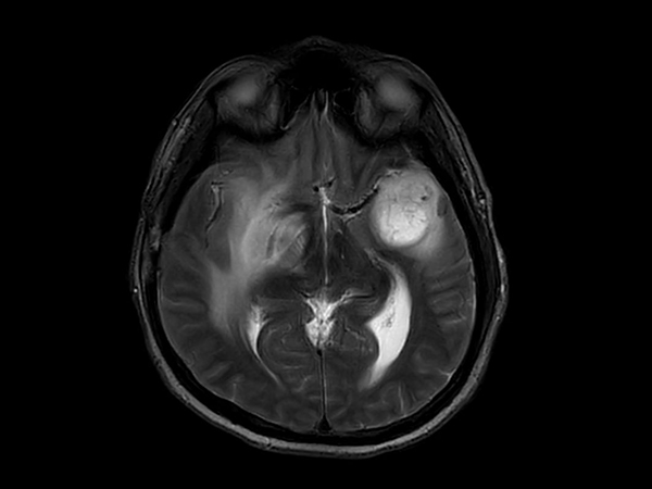
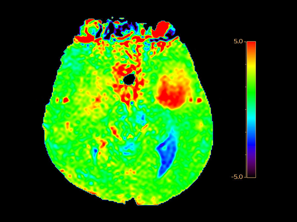
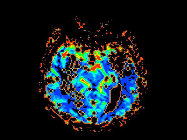
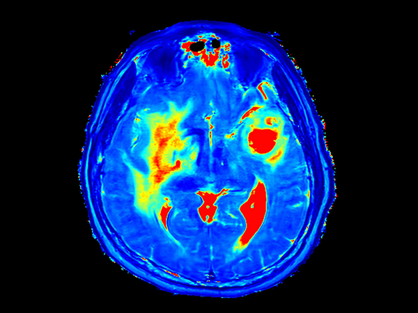
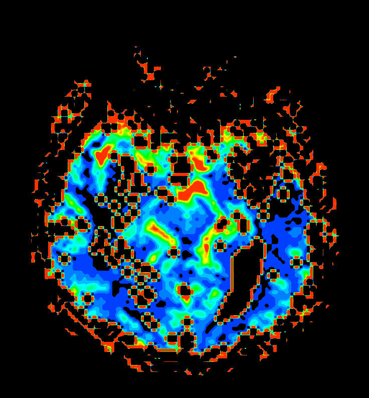
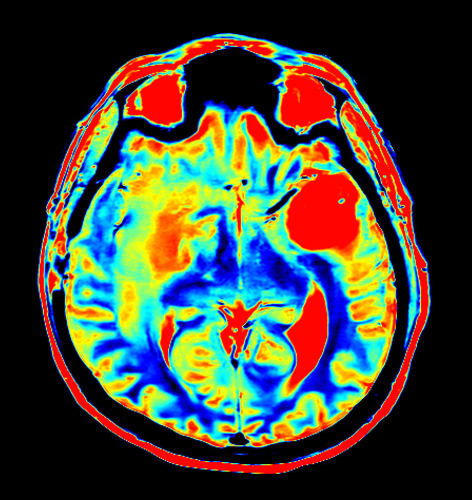
Axial T2* Perfusion (TTP)
![Axial T2* Perfusion (TTP)]()
-
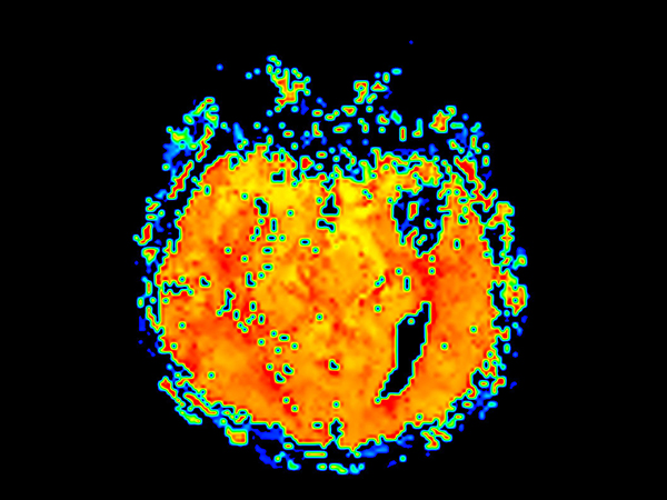
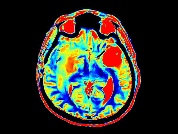
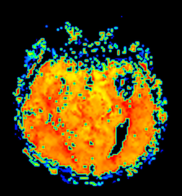
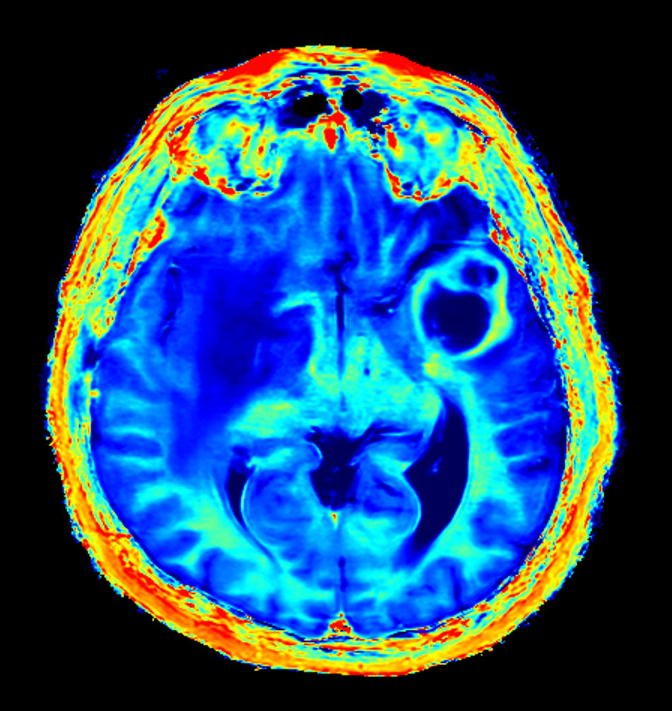
Axial T2* Perfusion (NI)
![Axial T2* Perfusion (NI)]()
-
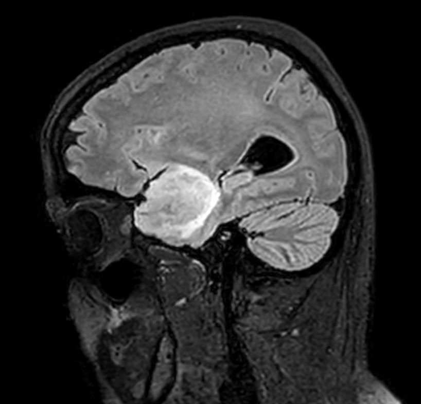
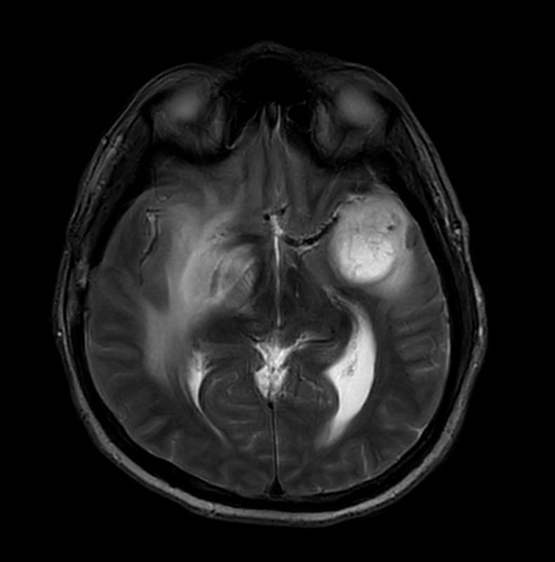
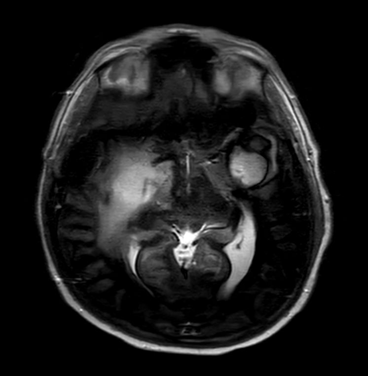
Sagittal 3D T1w TFEPost-Gado
![Sagittal 3D T1w TFE<b>Post-Gado</b>]()
-
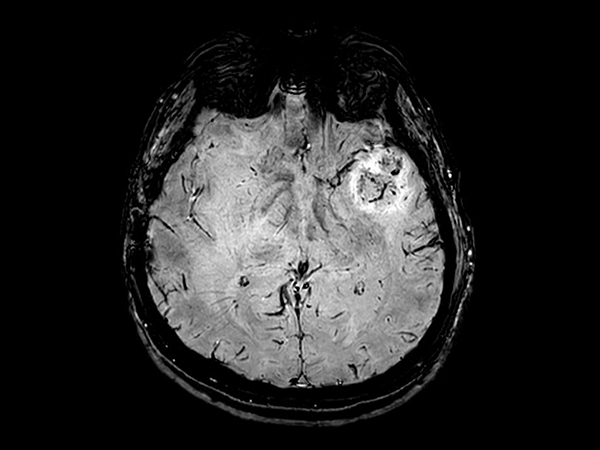
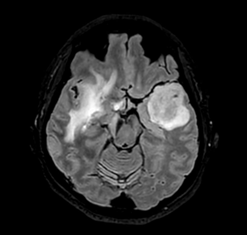
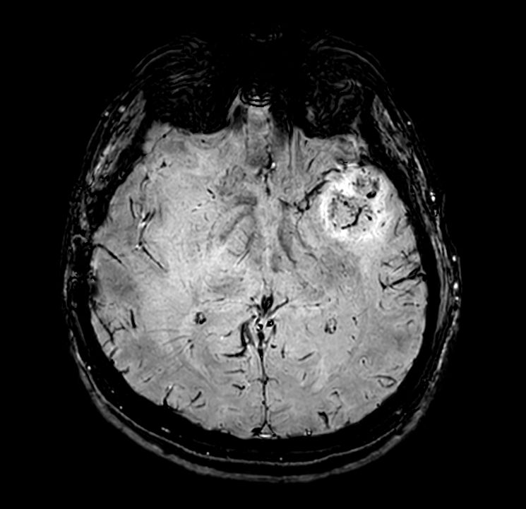
3D T1w TFE (Axial reformat)Post-Gado
![3D T1w TFE (Axial reformat)<b>Post-Gado</b>]()
-
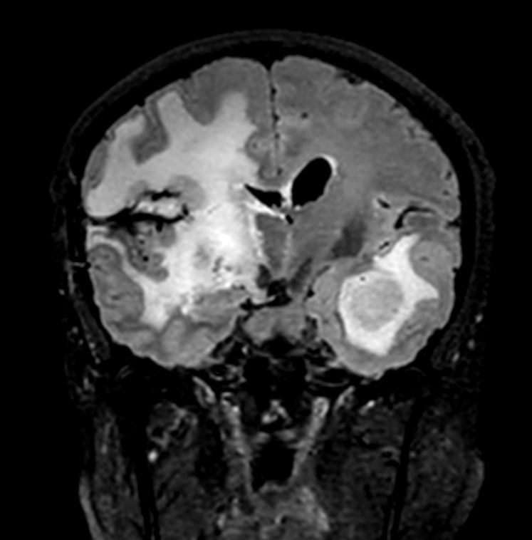
3D T1w TFE (Coronal reformat)Post-Gado
![3D T1w TFE (Coronal reformat)<b>Post-Gado</b>]()
-
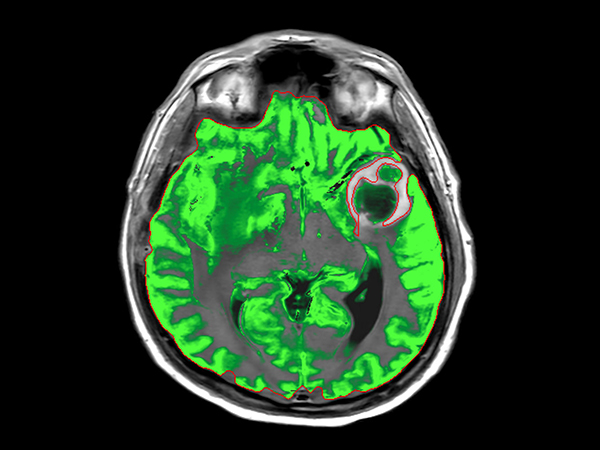
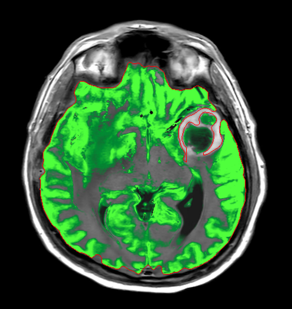
Axial SyntAc
![Axial SyntAc]()
-
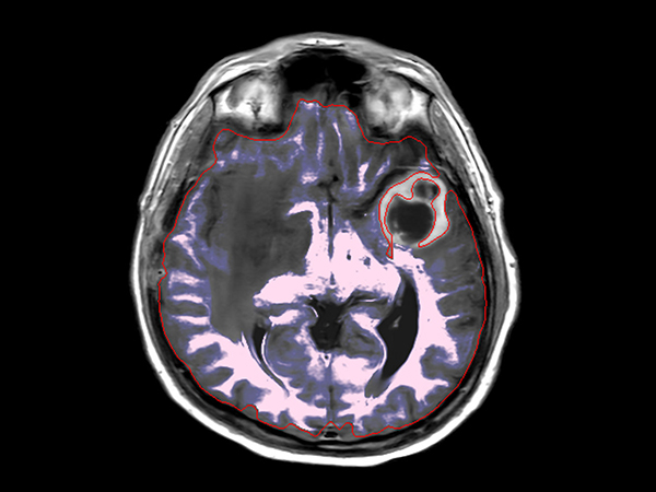
Axial SyntAc (Grey Matter)
![Axial SyntAc (Grey Matter)]()
-
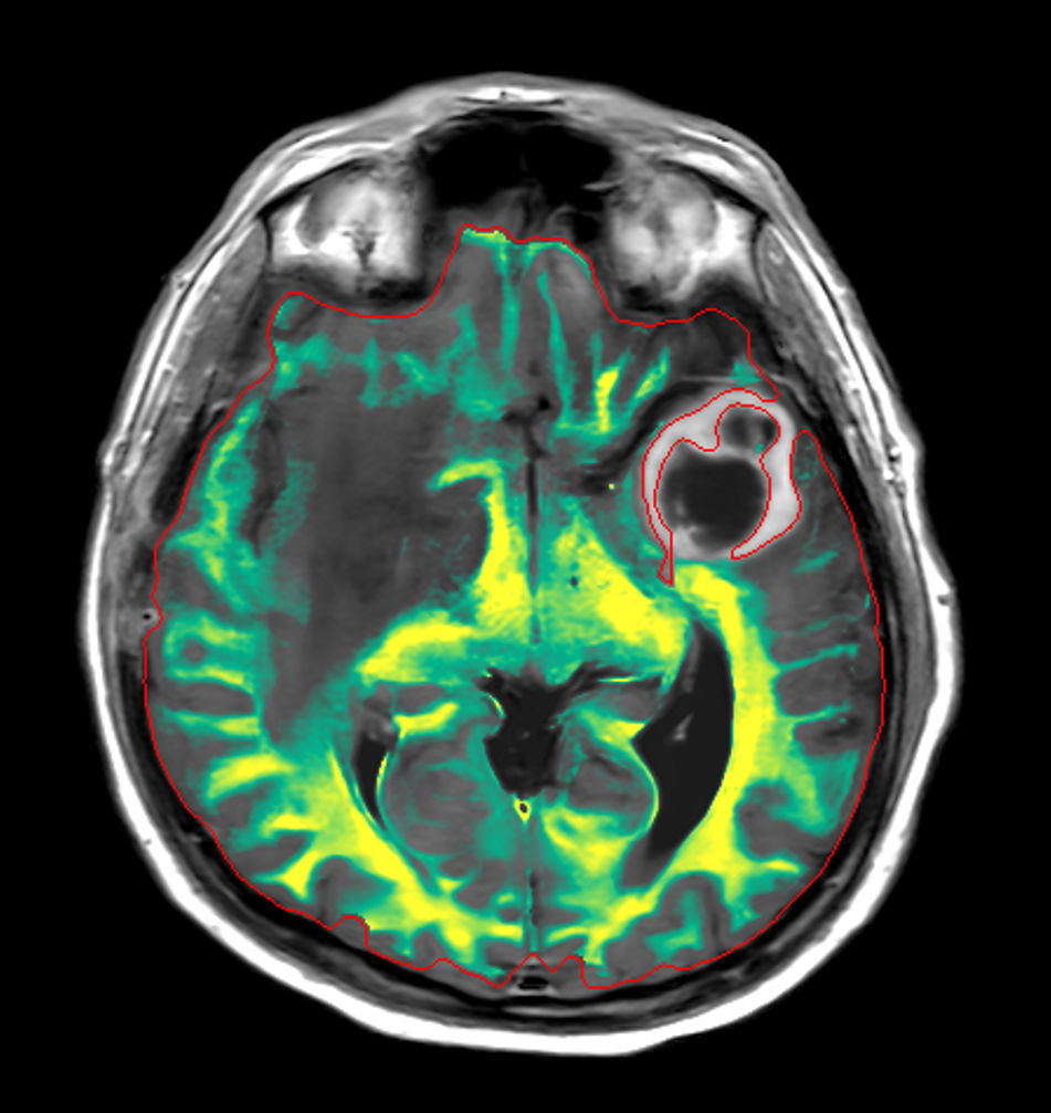
Axial SyntAc (White Matter)
![Axial SyntAc (White Matter)]()
-
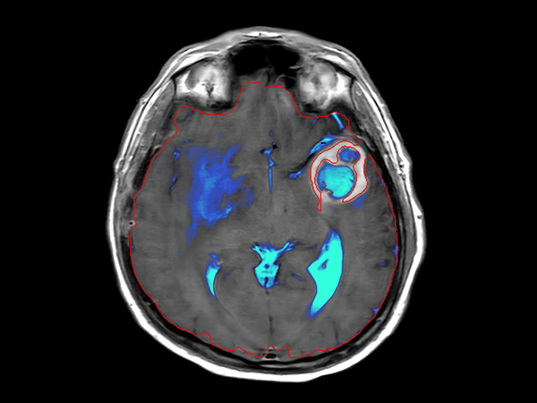
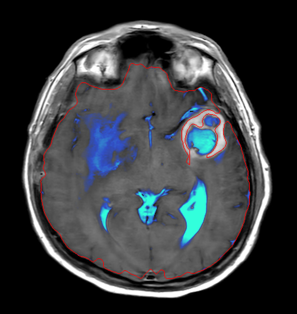
Axial SyntAc (CSF)
![Axial SyntAc (CSF)]()
-

Axial SyntAc (No GM, WM, CSF)
![Axial SyntAc (No GM, WM, CSF)]()
-
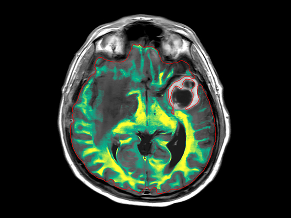
Axial SyntAc (Myelin)
![Axial SyntAc (Myelin)]()
-
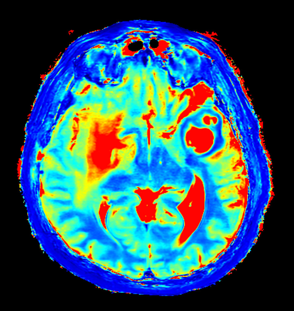
Axial SyntAc (T1 Map)
![Axial SyntAc (T1 Map)]()
-
Axial SyntAc (T2 Map)
![Axial SyntAc (T2 Map)]()
-
Axial SyntAc (PD Map)
![Axial SyntAc (PD Map)]()
-
Axial SyntAc (R1 Map)
![Axial SyntAc (R1 Map)]()
-
Axial SyntAc (R2 Map)
![Axial SyntAc (R2 Map)]()


Download ExamCards
Large lesion brain imaging with synthetic MRIMore Information
- Web page: No compromise. Image quality and speed at your fingertips.
- FieldStrength video and article: Discover how Mermaid Beach has shortened exam times and improved image quality in MR
- Customer webinar: Enhancing Diagnostic Confidence with Philips SmartSpeed at the University Clinic Bonn
- FieldStrength article: Scan fast and have more time for advanced MRI techniques
- FieldStrength article: UBC researchers advance their multiple sclerosis imaging
- Video testimonial: Gain time for advanced neuro MRI
- Webinar: Advancements in 3.0T imaging by team of radiologists from Technical University of Munich
- Webinar: Improvements in clinical MR Neuro imaging by Joshua P Nickerson, MD
- White paper: Philips SmartSpeed
- Brochure: MR Workspace and clinical applications portfolio
*SyMRI NEURO, Synthetic MR, AB, Sweden
Results from case studies are not predictive of results in other cases. Results in other cases may vary.