Go back
High quality, fast brain aneurysm imaging
Patient information
High quality, fast 3.0T imaging for brain aneurysm on the uniquely designed Philips MR 7700 imaging system. Advanced 3D TSE imaging technique is used for T2w and T1w (pre- and post-gado injection), to acquire high resolution data in multiple directions in only one single scan. Up to 35% higher SNR is achieved for echo-planar (EPI) diffusion imaging thanks to the added power of the XP gradients. SWIp is added to provide high resolution 3D susceptibility weighted brain imaging including phase maps to support advanced diagnosis.Gallery
-
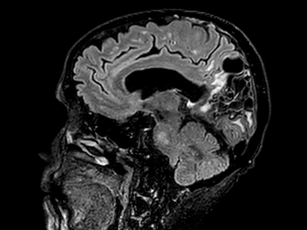
Sagittal 3D VIEW - T2w FLAIR
![Sagittal 3D VIEW - T2w FLAIR]()
-
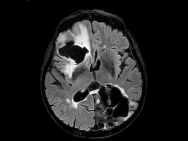
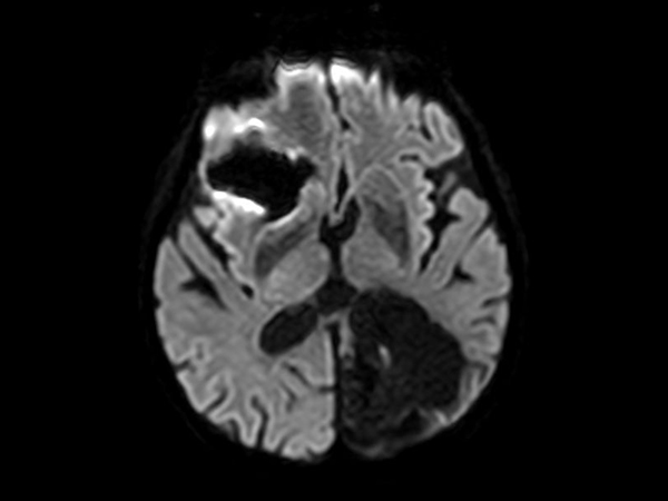
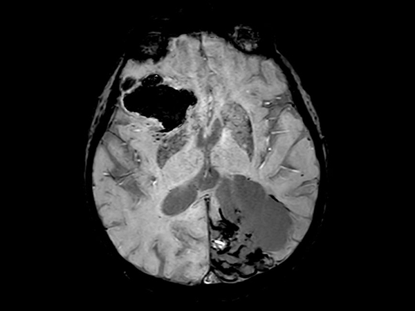
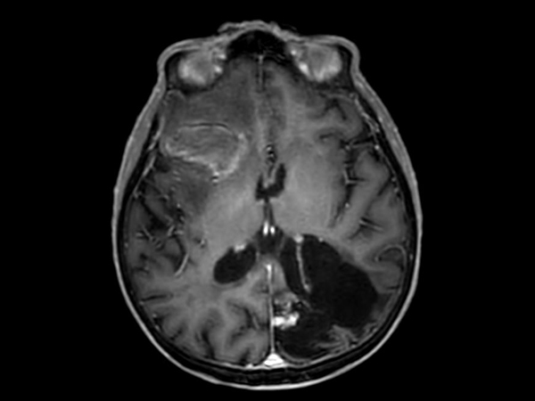
3D VIEW - T2w FLAIR (axial reformat)
![3D VIEW - T2w FLAIR (axial reformat)]()
-
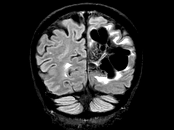
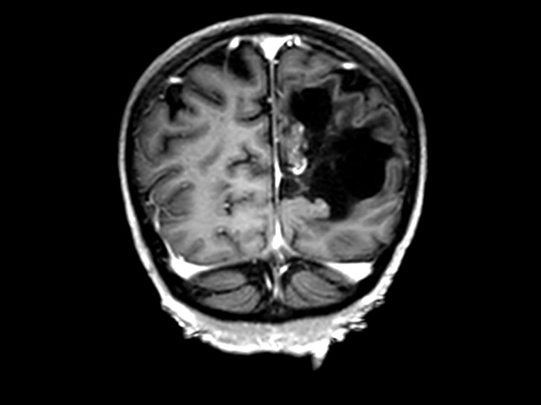
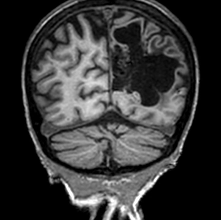
3D VIEW - T2w FLAIR (coronal reformat)
![3D VIEW - T2w FLAIR (coronal reformat)]()
-
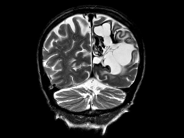
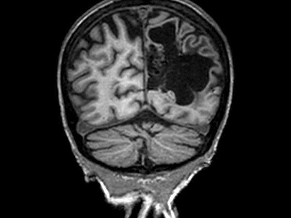
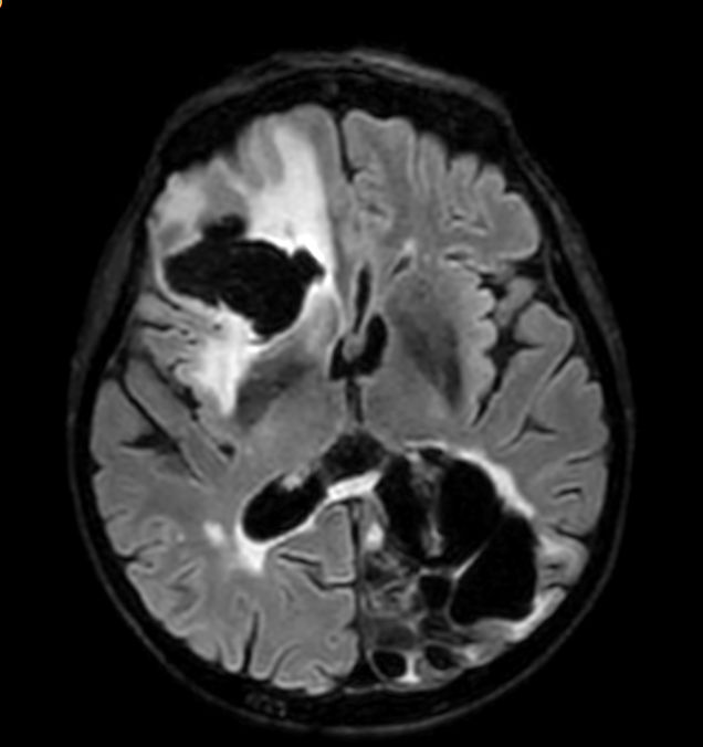
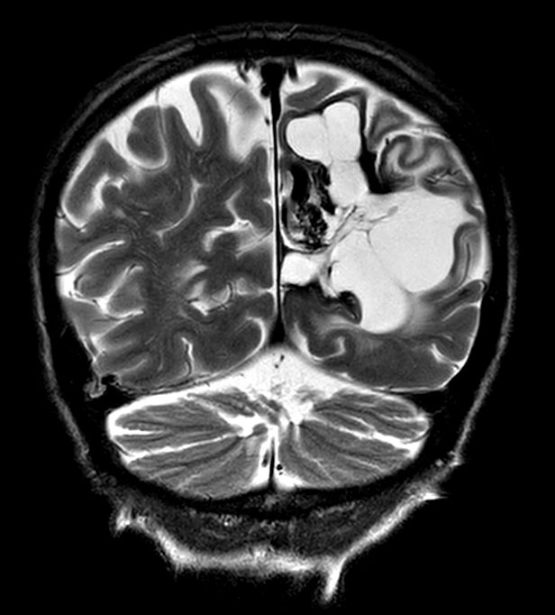
Coronal T2w TSE
![Coronal T2w TSE]()
-
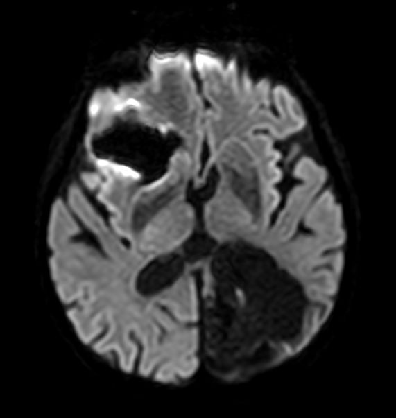
Axial Diffusion (b1000)
![Axial Diffusion (b1000)]()
-
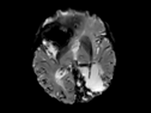
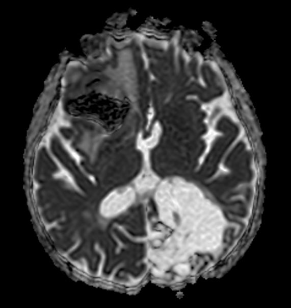
Axial Diffusion (ADC)
![Axial Diffusion (ADC)]()
-
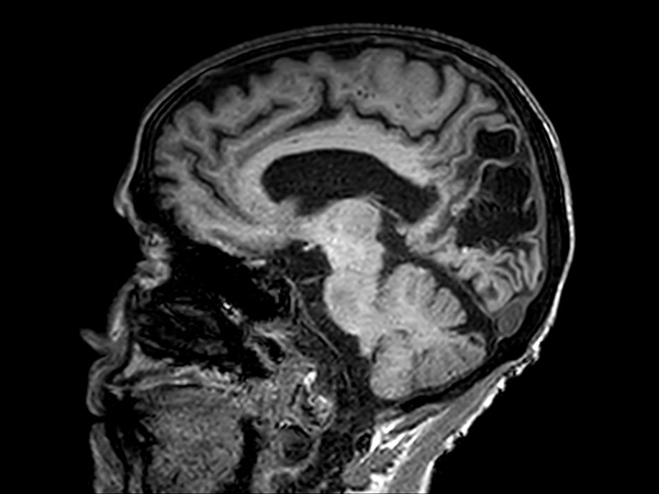
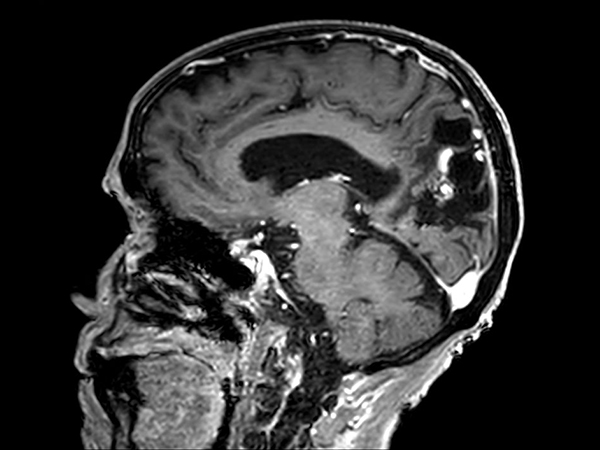
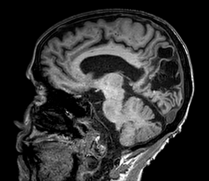
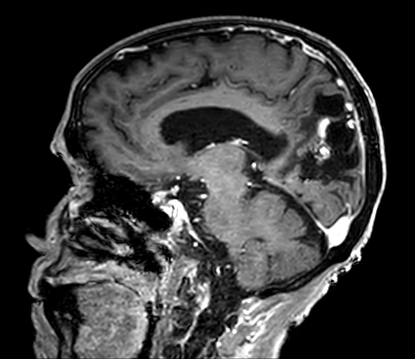
Sagittal 3D T1w FFE
![Sagittal 3D T1w FFE]()
-
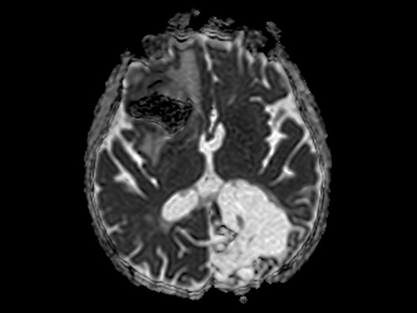
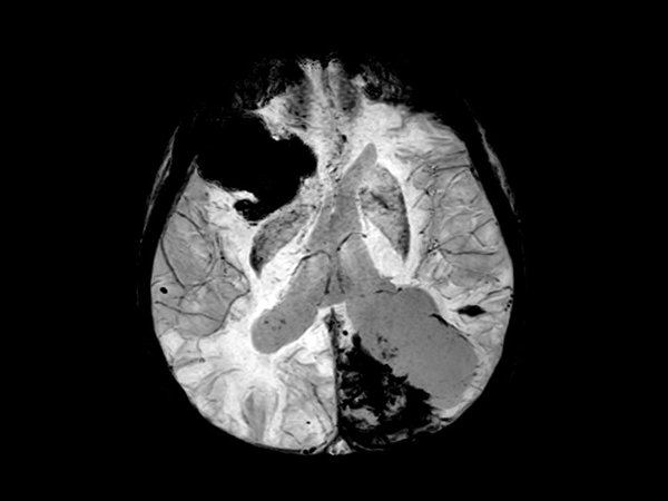
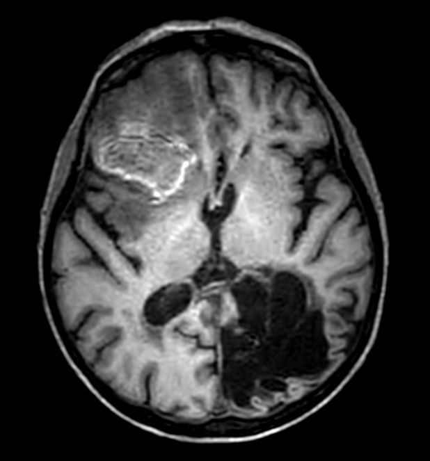
3D T1w FFE (axial reformat)
![3D T1w FFE (axial reformat)]()
-
3D T1w FFE (coronal reformat)
![3D T1w FFE (coronal reformat)]()
-
Axial SWIp (Modulus)
![Axial SWIp (Modulus)]()
-
Axial SWIp (minIP)
![Axial SWIp (minIP)]()
-
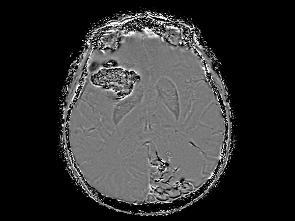
Axial SWIp (Phase)
![Axial SWIp (Phase)]()
-
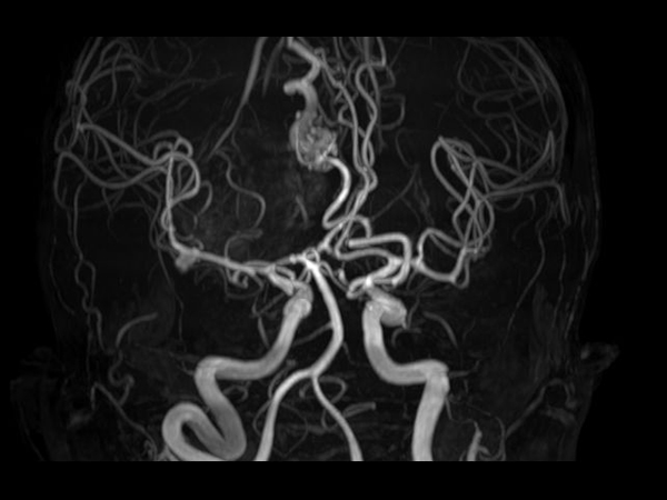
Angio (Time-of-Flight)
-
Perfusion (100 dynamics)
-
Sagittal 3D T1w FFE (subtraction)
![Sagittal 3D T1w FFE (subtraction)]()
-
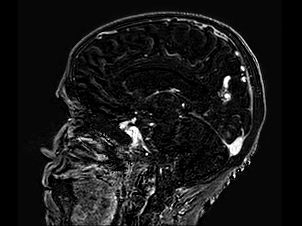
Sagittal 3D T1w FFE +gado
![Sagittal 3D T1w FFE +gado]()
-
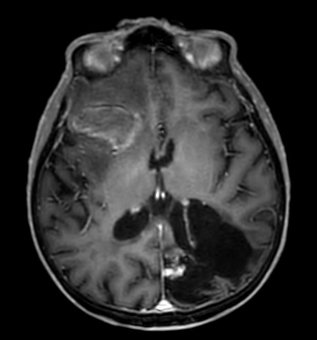
3D T1w FFE (axial reformat) +gado
![3D T1w FFE (axial reformat) +gado]()
-
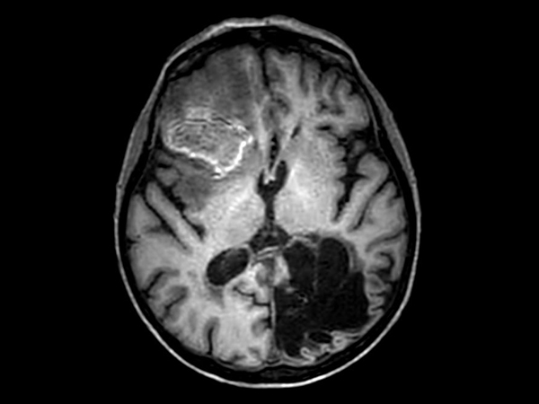
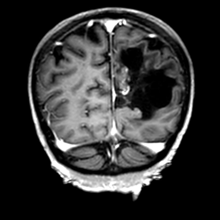
3D T1w FFE (coronal reformat) +gado
![3D T1w FFE (coronal reformat) +gado]()


Download ExamCards
High quality, fast brain aneurysm imagingMore Information
- FieldStrength article: High-powered gradients boost MR research projects at UKM
- Testimonial video: Unmatched performance and precision at Tufts Medical Center Boston
- Testimonial video: Clinical value of MR 7700 in MSK imaging at Tufts Medical Center Boston
- Customer webinar: Experiences with MR7700 at University Hospital Brest
- Testimonial video: Enhancing patient comfort at Tufts Medical Center Boston
- FieldStrength article: Scan fast and have more time for advanced MRI techniques
- Video testimonial: Gain time for advanced neuro MRI
- Video testimonial: Clinical use of SWIp for neuro imaging
- Webinar: Advancements in 3.0T imaging by team of radiologists from Technical University of Munich
- Webinar: Improvements in clinical MR Neuro imaging by Joshua P Nickerson, MD
- Webinar: Advanced Neuro MR applications by Jan W. Casselman, MD, PhD
- Brochure: MR Workspace and clinical applications portfolio
*Results from case studies are not predictive of results in other cases. Results in other cases may vary.