Go back
Multi Nuclei - Dynamic calf muscle imaging (31P)
Patient information
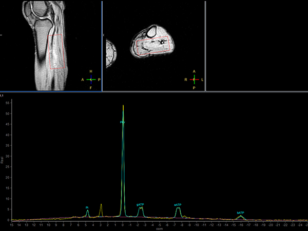
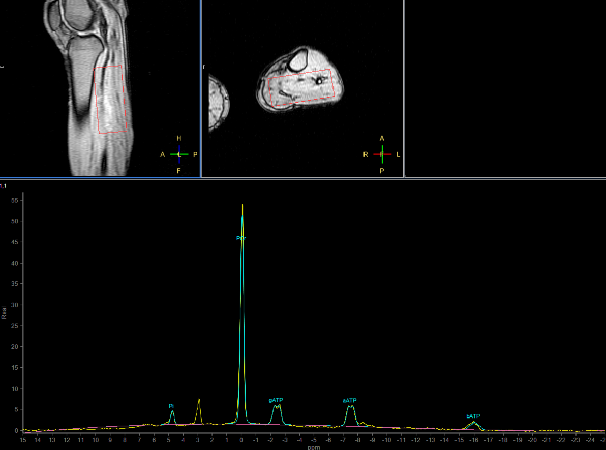
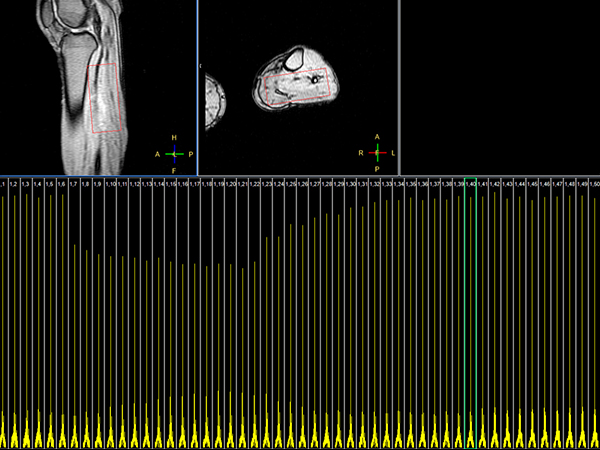
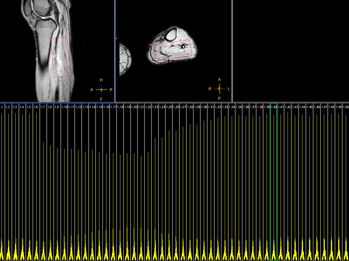
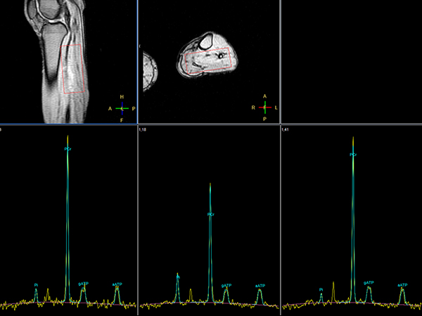
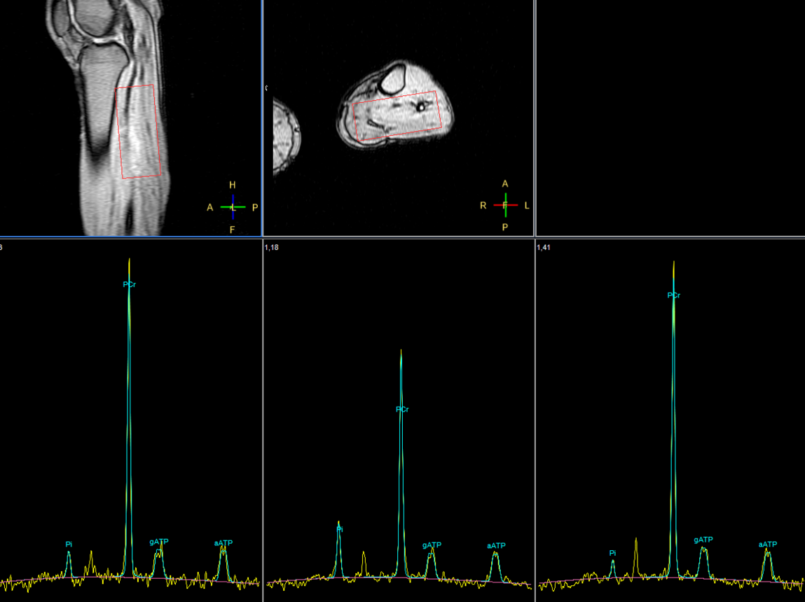
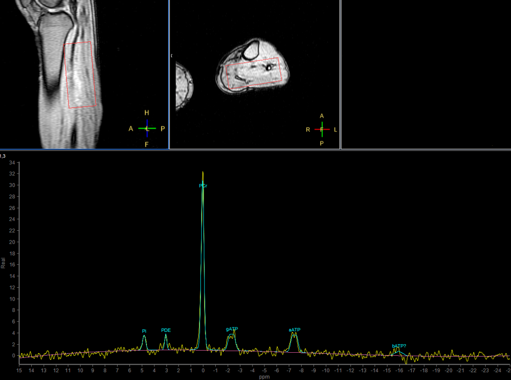
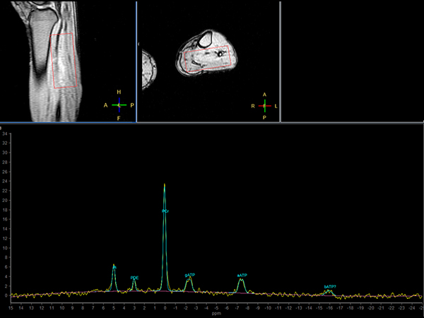
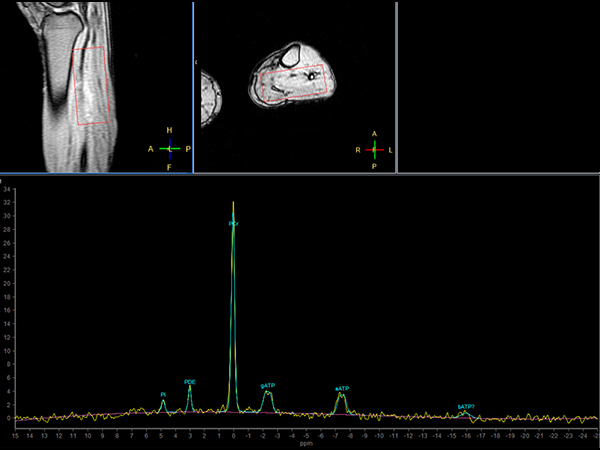
Make multi-nuclei imaging and spectroscopy become part of your clinical operations via a seamless integrated workflow for multi-nuclei image acquisition, spectroscopy, reconstruction, and viewing. The nucleus is just a scan parameter like any other sequence parameter. A single ExamCard can be used to run both proton and non-proton imaging. Reconstruction and viewing of non-proton images or spectra, as well as the process for sending the data to PACS is fully integrated, so workflow does not differ from proton imaging. The transmit-receive 31P flex coil, with a 14 cm diameter, is immediately recognized by the ExamCard interface. Improved SNR and simplified spectra* are achieved by combining body coil decoupling with the transmit-receive surface coils. This is an example of dynamic 31P spectroscopy showing how the metabolism (signals of PCr and PI) change during a calf-muscle exercise.Gallery


Download ExamCards
Multi Nuclei - Dynamic Muscle imaging (31P)More Information
- Brochure: Multi Nuclei
- Web page: Seamless integration of multi-nuclei
- Video: It’s not just confidence. It’s precision diagnosis
*Results from case studies are not predictive of results in other cases. Results in other cases may vary.