Orbit with melanoma
Patient information
Patient with uveal melanoma referred for orbital MR imaging. Until about 5 years ago, 2D ultrasound imaging was the gold standard. Now MRI offers the opportunity for 3D imaging along with different weightings for the radiologist to provide a confident diagnosis.4 different 3D isotropic sequences, T1W with and without fat saturation, T2W and T1W fat saturation post-contrast are used to evaluate the tumor prior to treatment. Isotropic sequences are used to allow for good quality multiplanar reconstruction corresponding to the location of the tumor; as the tumor can be located anywhere in the eye, optimal visualization often requires an assessment of different oblique planes, which is facilitated by the 3D isotropic sequences. The pre-contrast sequences help evaluate the tumor size and location and the post-contrast sequence provides additional information such as identifying tumor that has seeped through the sclera behind the eye and differentiating tumor vs associated retinal detachment.
Higher in-plane resolution 2D sequences are used to better evaluate the tumor and screen for infiltration to the adjacent structures, which can have direct therapeutic consequences.
Perfusion and diffusion scans are used for differential diagnosis. The diffusion sequence can help a radiologist confirm whether a tumor is malignant or not and the perfusion results can show the effects of treatment. Using ultrasound to evaluate the effects of treatment takes longer due to post-treatment inflammation.
Gallery
-

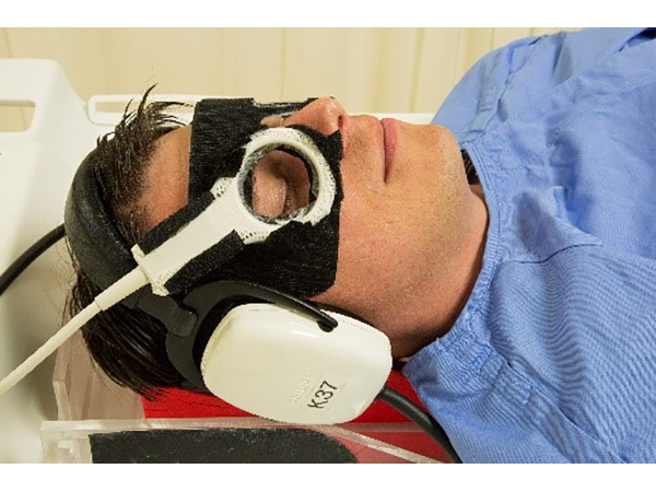
Positioning of ds Micro 47 coil.
Note: Goggles illustrated are not Philips product.
![Positioning of ds Micro 47 coil.<p>Note: Goggles illustrated are not Philips product.]()
-
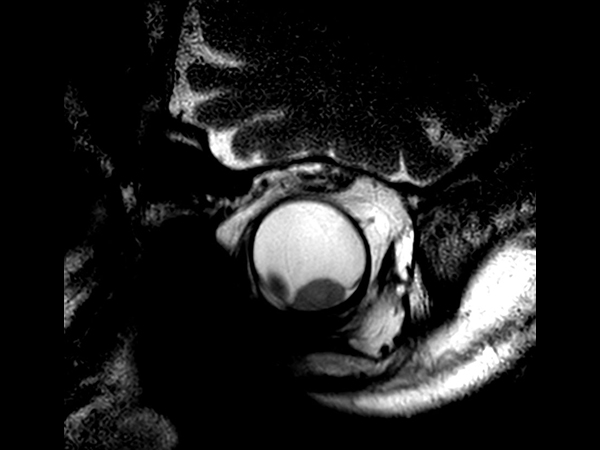
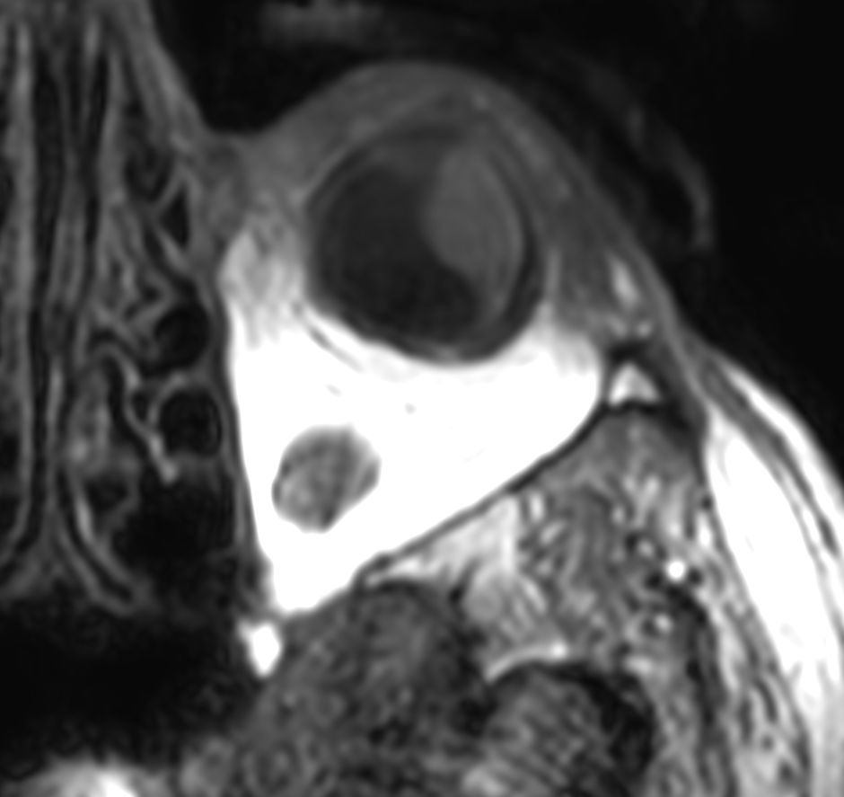
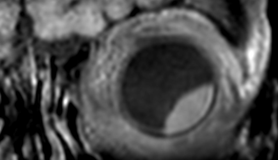
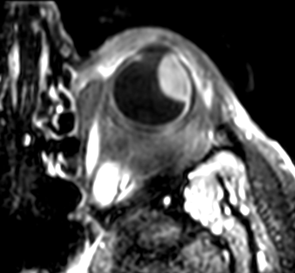
2D T2w TSE - Oblique coronal
![2D T2w TSE - Oblique coronal]()
-
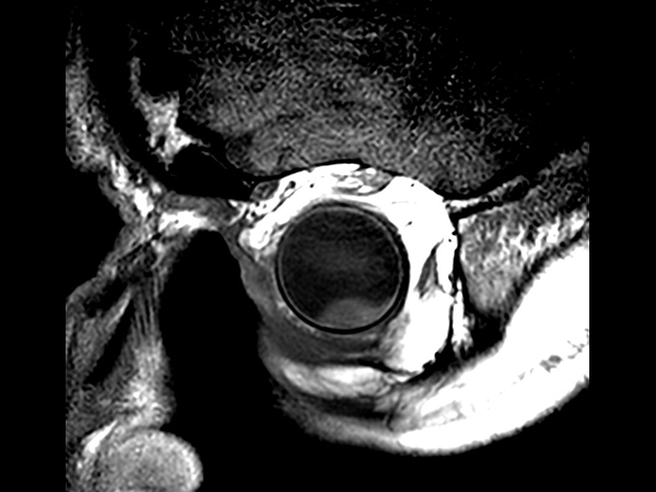
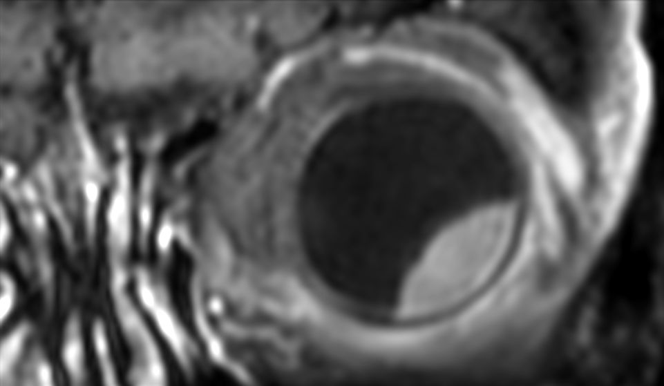
2D T1w TSE - Oblique coronal
![2D T1w TSE - Oblique coronal]()
-
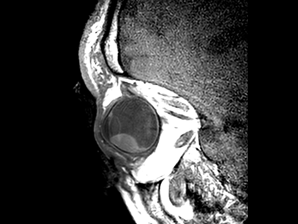
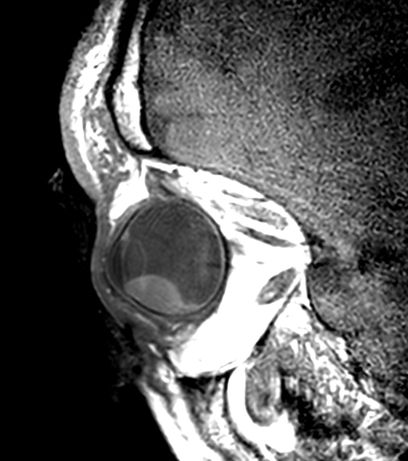
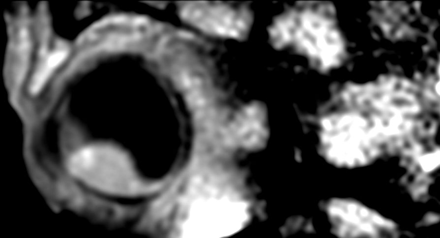
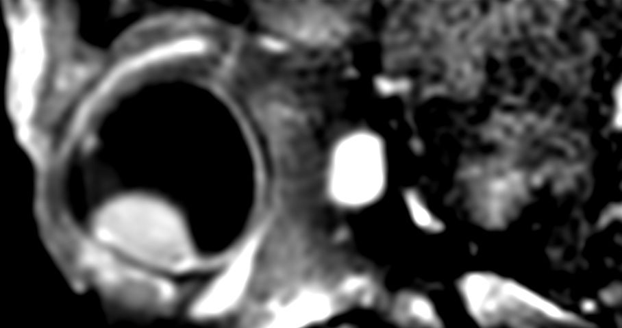
2D T1w TSE - Oblique sagittal
![2D T1w TSE - Oblique sagittal]()
-
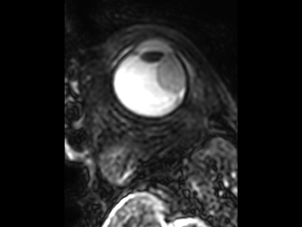
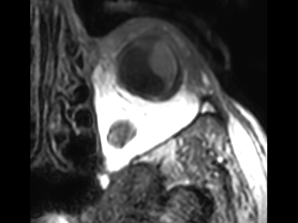
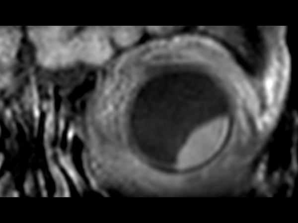
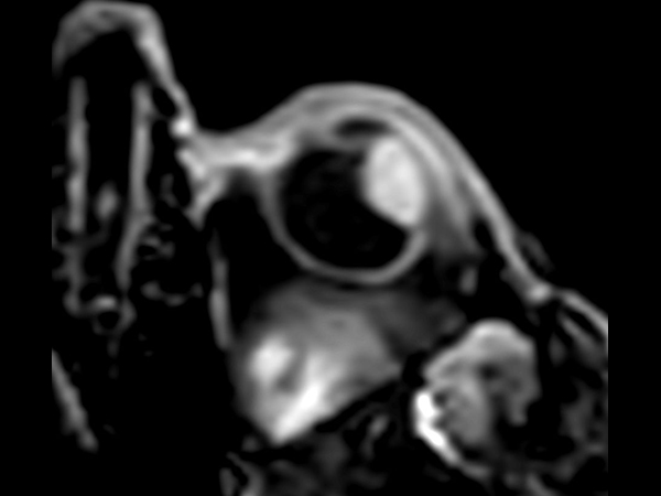
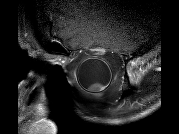
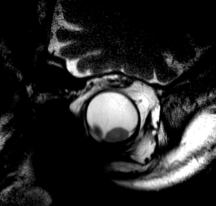
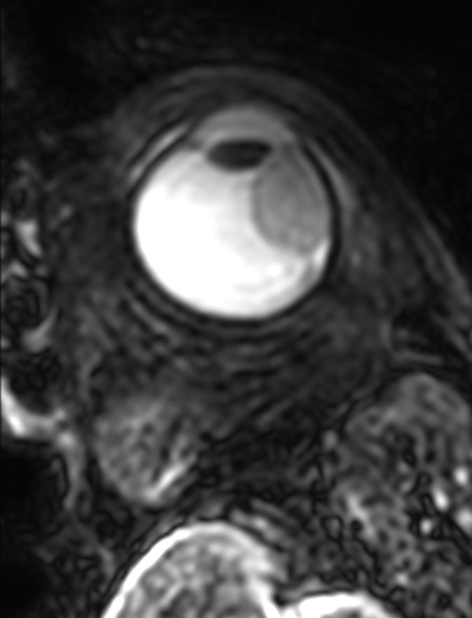
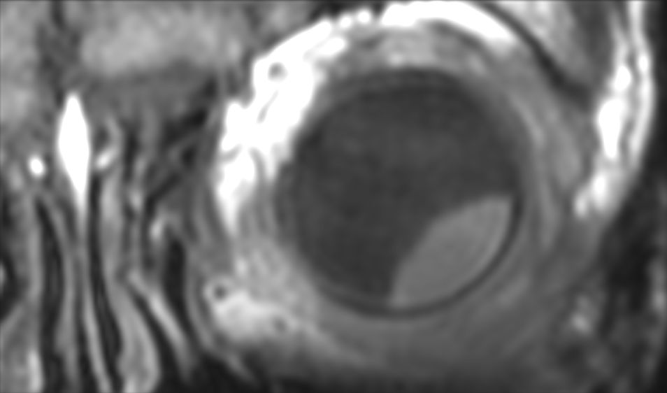
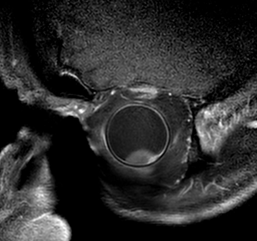
3D T2w TSE - Axial
![3D T2w TSE - Axial]()
-
3D T2w TSE - Coronal reformat
![3D T2w TSE - Coronal reformat]()
-
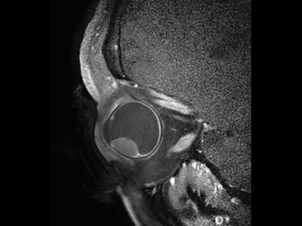
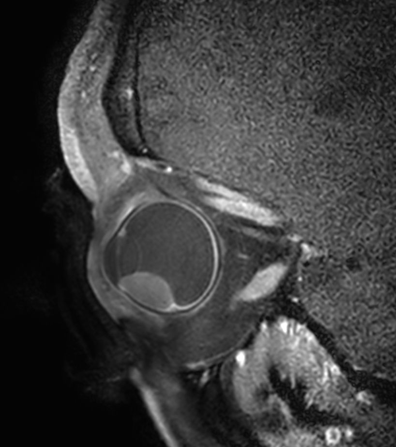
3D T2w TSE - Sagital reformat
![3D T2w TSE - Sagital reformat]()
-
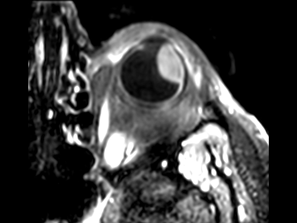
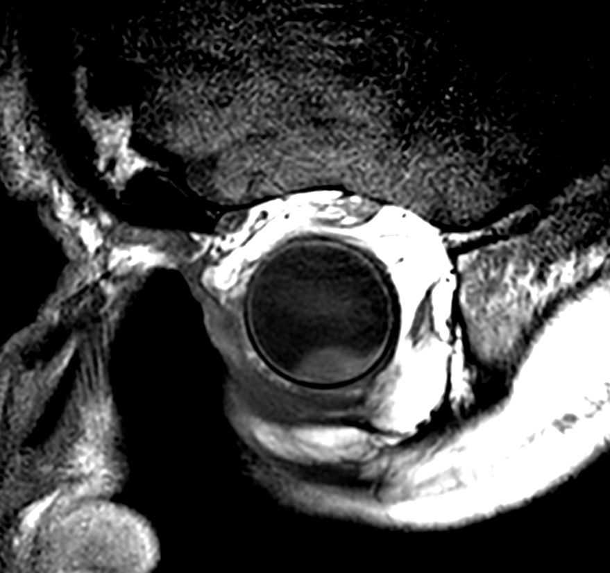
3D T1w TSE - Axial
![3D T1w TSE - Axial]()
-
3D T1w TSE - Coronal reformat
![3D T1w TSE - Coronal reformat]()
-
3D T1w TSE - Sagital reformat
![3D T1w TSE - Sagital reformat]()
-
3D T1w TSE FatSat - Axial
![3D T1w TSE FatSat - Axial]()
-
3D T1w TSE FatSat - Coronal reformat
![3D T1w TSE FatSat - Coronal reformat]()
-
3D T1w TSE FatSat - Sagital reformat
![3D T1w TSE FatSat - Sagital reformat]()
-
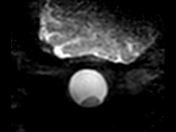
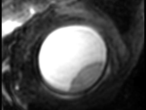
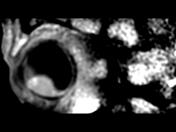
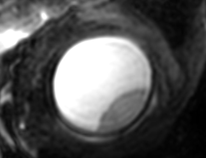
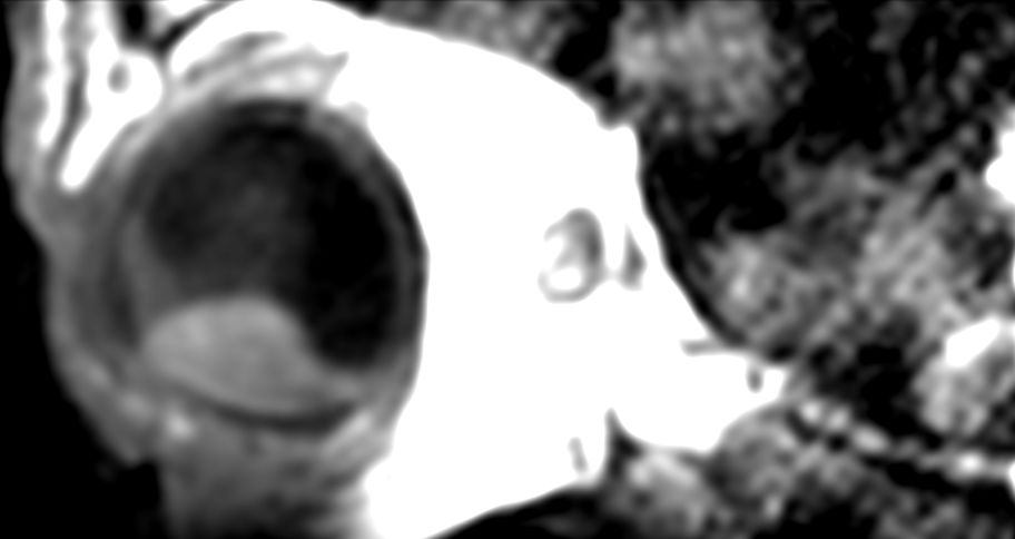
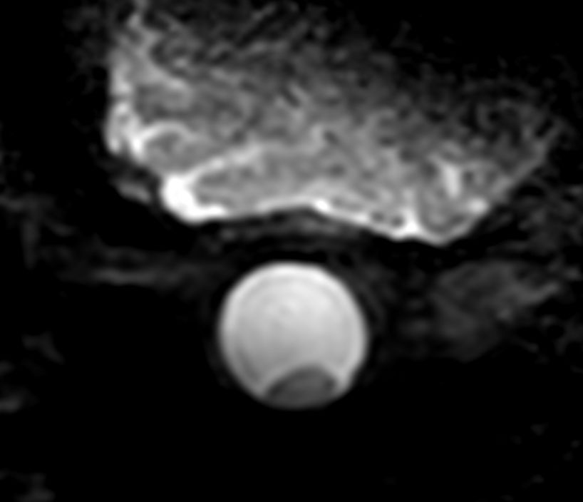
DWI TSE - b0
![DWI TSE - b0]()
-

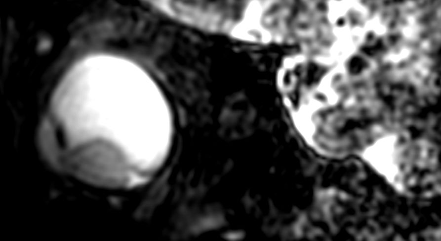
DWI TSE - b800
![DWI TSE - b800]()
-
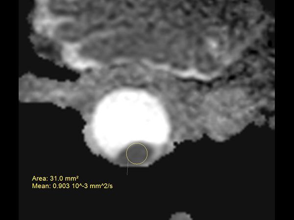
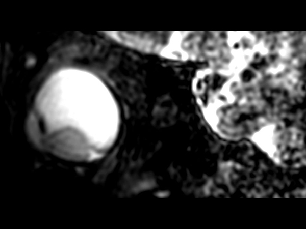
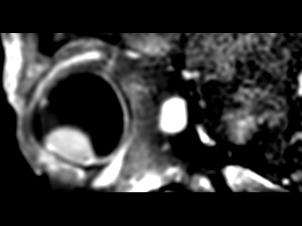
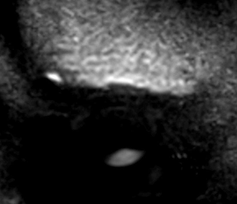
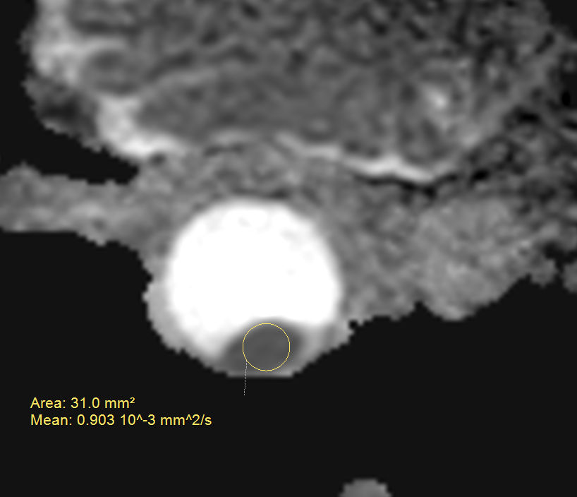
DWI TSE - ADC
![DWI TSE - ADC]()
-
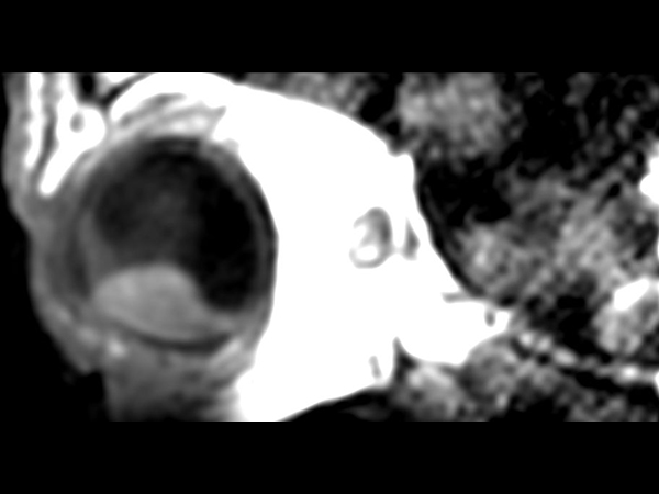
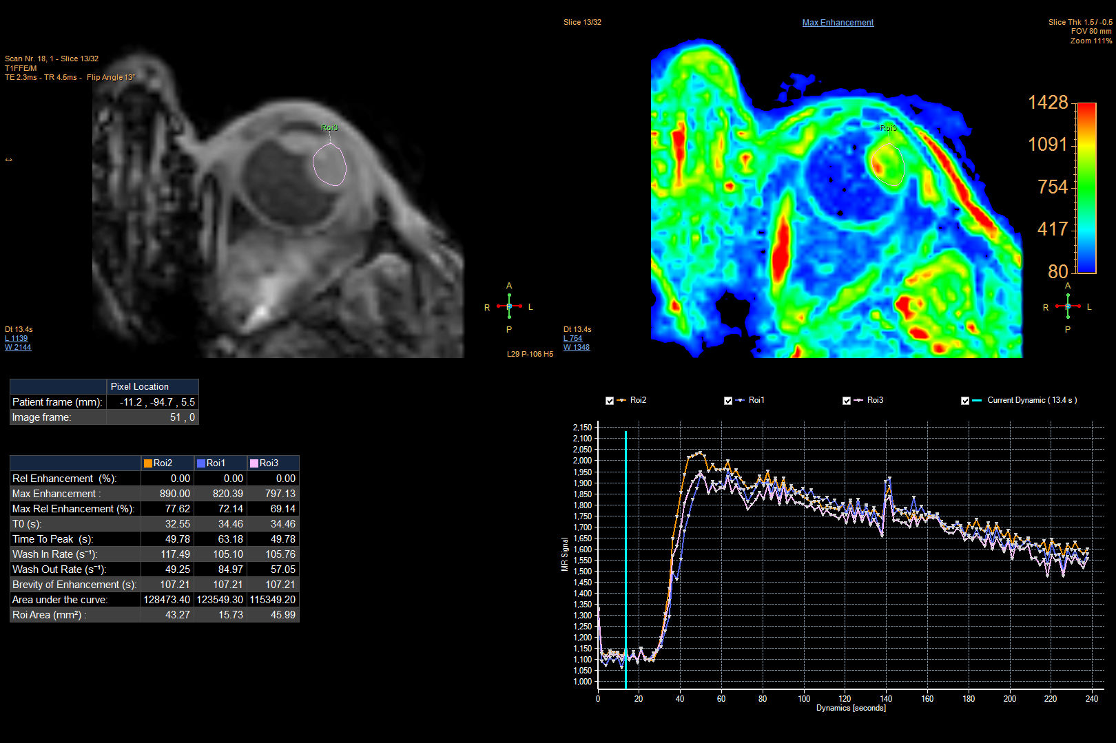
Perfusion - 125 dynamics
-
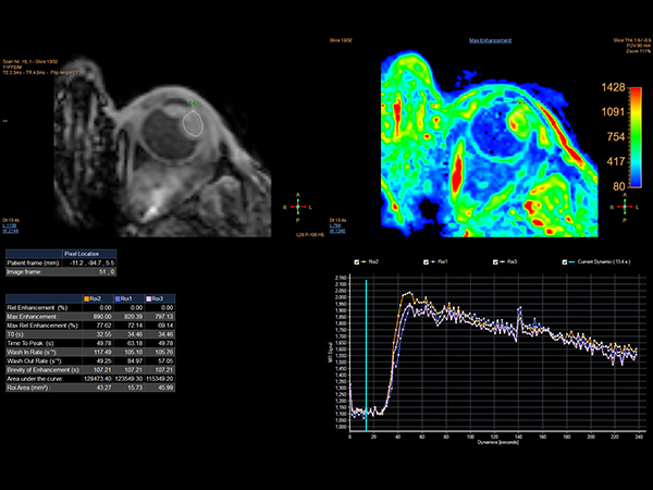
Perfusion - 125 dynamics (IntelliSpace Portal)
![Perfusion - 125 dynamics (IntelliSpace Portal)]()
-
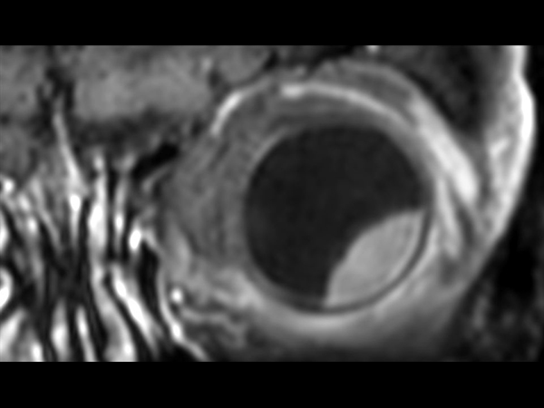
2D T1w TSE FatSat post gado - Oblique coronal
![2D T1w TSE FatSat post gado - Oblique coronal]()
-
2D T1w TSE FatSat post gado - Oblique sagital
![2D T1w TSE FatSat post gado - Oblique sagital]()
-
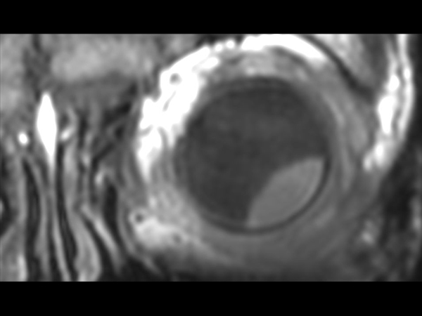
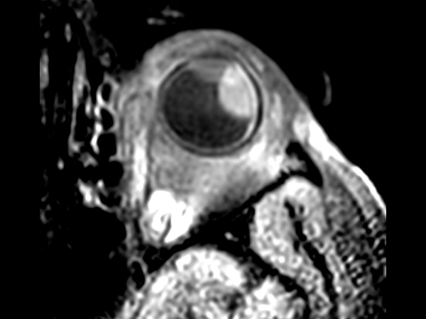
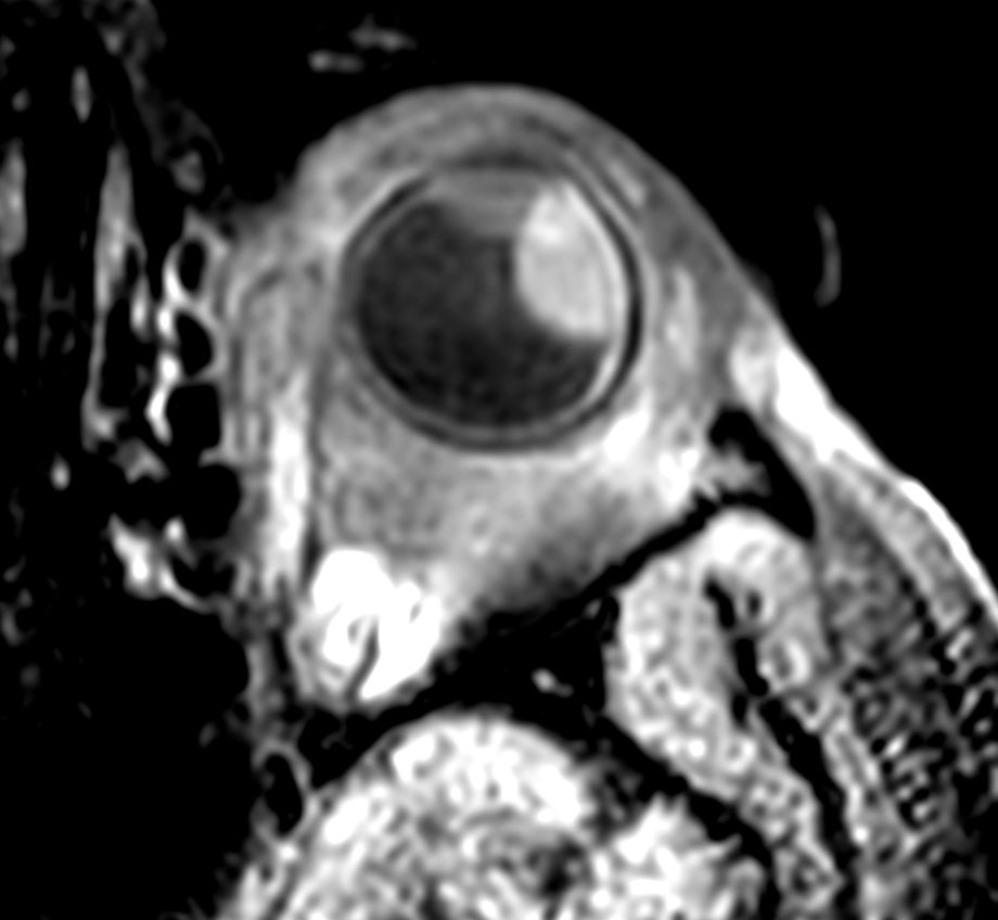
3D T1w TSE FatSat post gado - Axial
![3D T1w TSE FatSat post gado - Axial]()
-
3D T1w TSE FatSat post gado - Coronal reformat
![3D T1w TSE FatSat post gado - Coronal reformat]()
-
3D T1w TSE FatSat post gado - Sagital reformat
![3D T1w TSE FatSat post gado - Sagital reformat]()
Download ExamCards
Orbit with melanomaMore Information
- FieldStrength article: Helping the multitasking MR technologist
- FieldStrength article: Scan fast and have more time for advanced MRI techniques
- FieldStrength article: Relaxed patients, reduced motion, improved productivity
- Video testimonial: Optimizing MRI workflow and patient comfort
- Webinar: Increasing MR Operational Efficiency with 3.0T by RWJ Barnabas Health Ambulatory Care Center
- Webinar: Advancements in 3.0T imaging by team of radiologists from Technical University of Munich
*Results from case studies are not predictive of results in other cases. Results in other cases may vary.