Go back
FLAIR* protocol for Multiple Sclerosis
Patient information
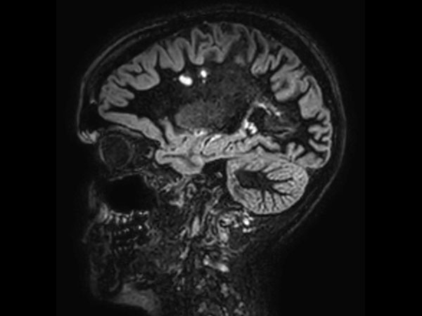
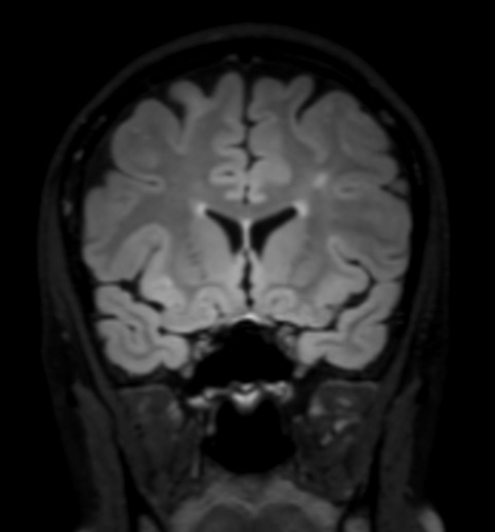
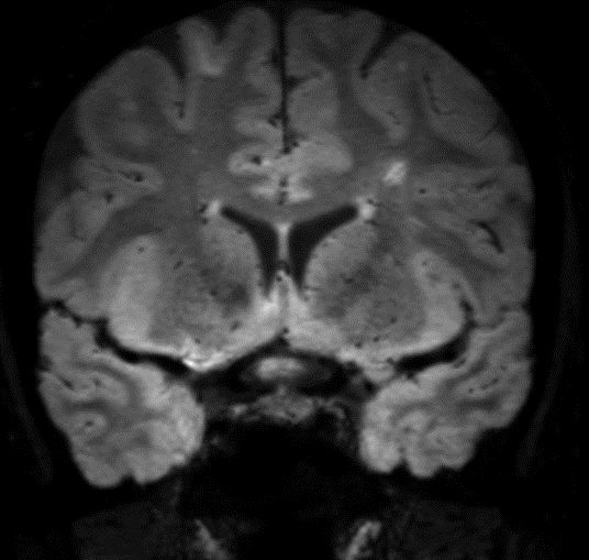
SWI sequence has a high sensitivity to enhance contrast for deoxygenated (venous) blood or calcium deposits. This may help, when used in combination with other clinical information, in the diagnosis of various neurological pathologies. 3D imaging lets you acquire high resolution data in multiple directions in one scan. Isotropic voxel size enables reformats in any plane without loss of resolution. FLAIR* requires offline post processing combining the contrast of 3D FLAIR and 3D SWI EPI into a single image. This enables the visualization of Central Vein Sign, mapping subcortical veins onto 3D FLAIR contrast images.Gallery
-
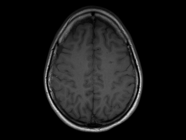
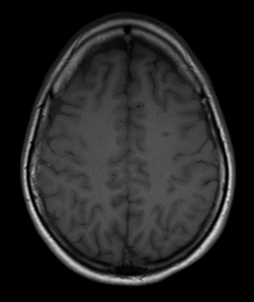
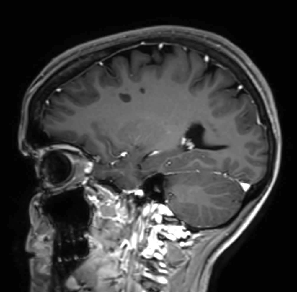
T1w SE
![T1w SE]()
-
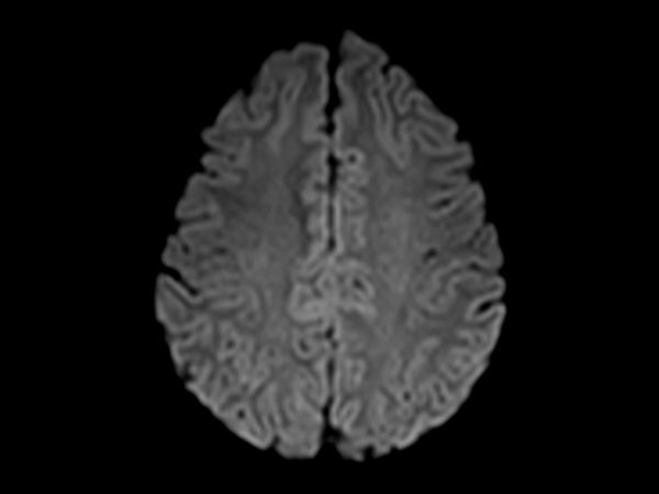
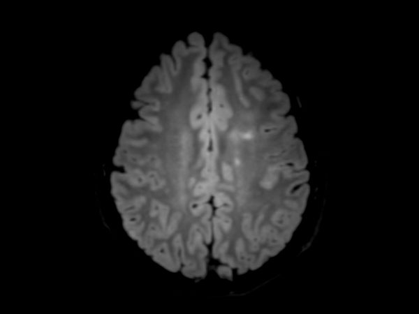
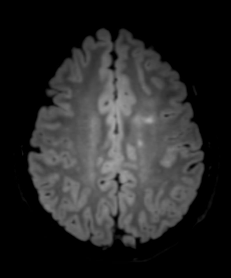
DWI b1000
![DWI b1000]()
-
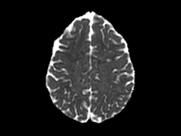
DWI b1000 (ADC)
![DWI b1000 (ADC)]()
-
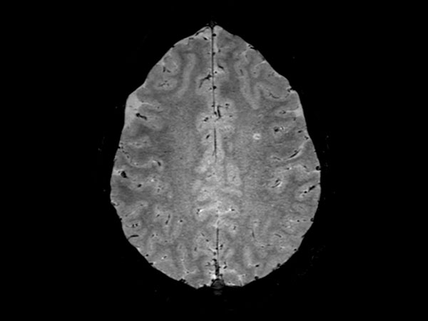
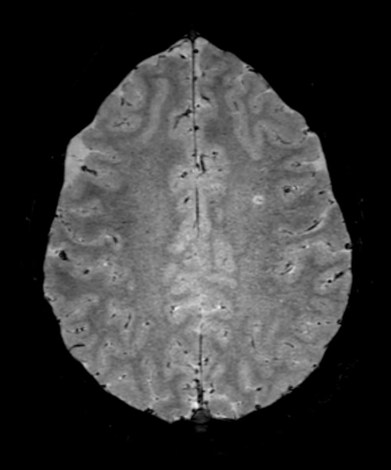
3D SWI EPI (reformat)
![3D SWI EPI (reformat)]()
-
3D SWI EPI (reformat)
![3D SWI EPI (reformat)]()
-
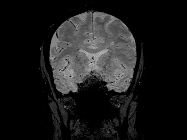
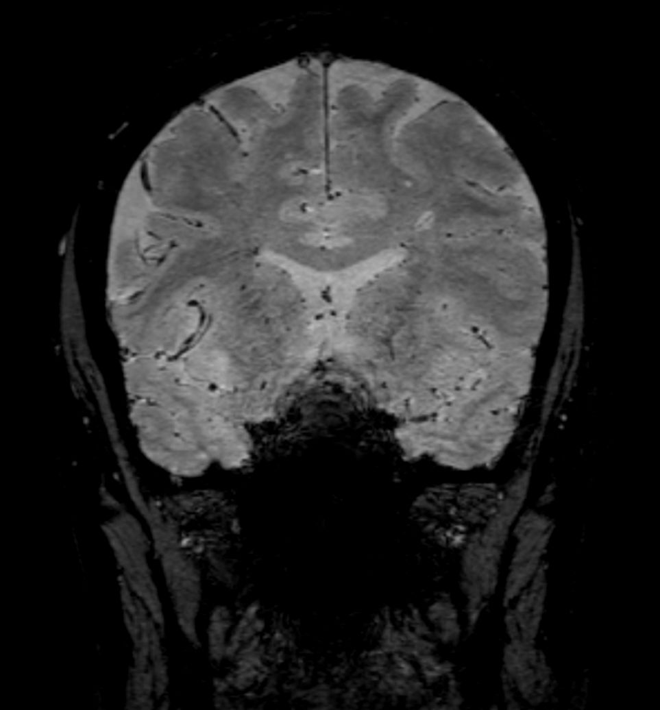
3D SWI EPI
![3D SWI EPI]()
-
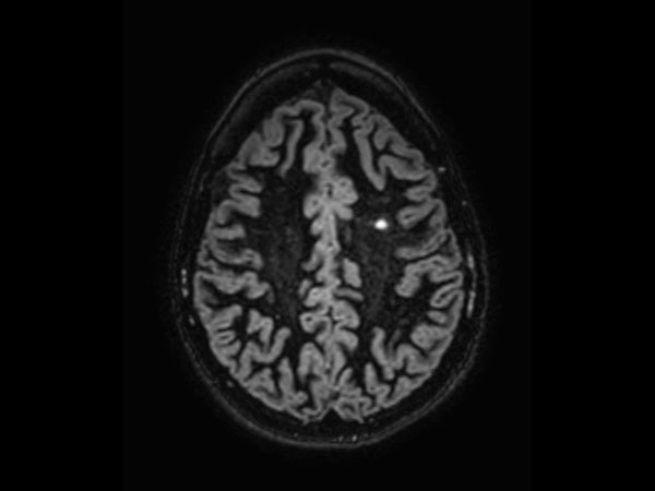
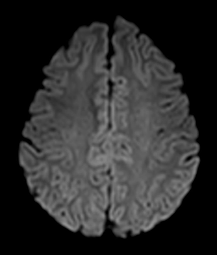
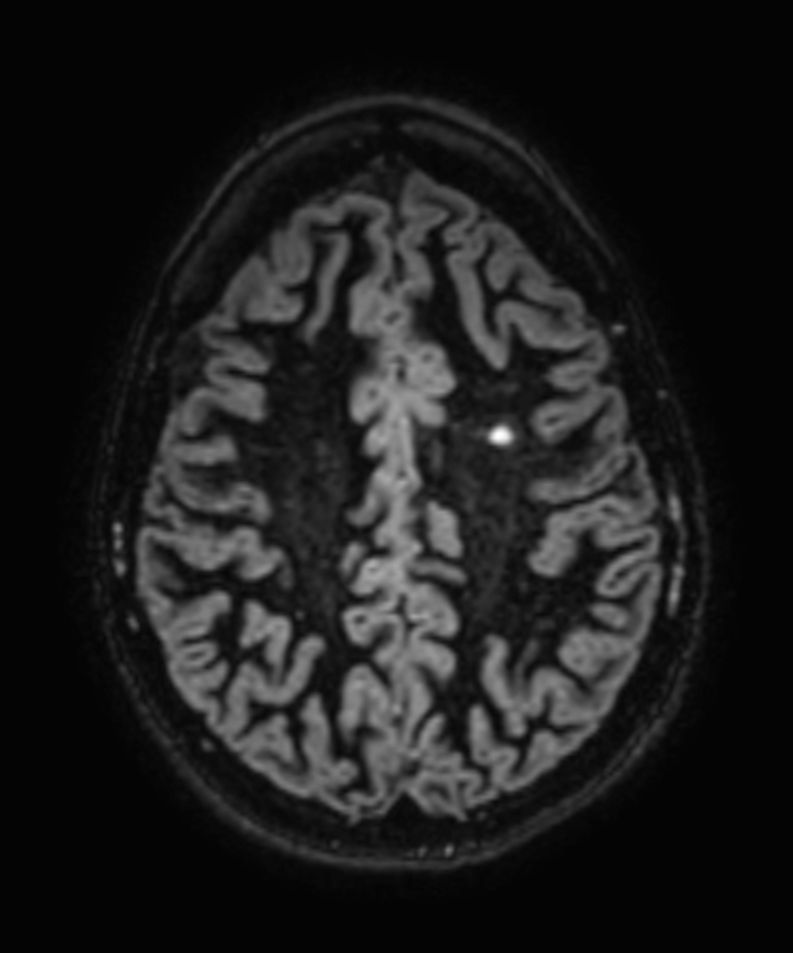
3D T2w FLAIR (reformat)
![3D T2w FLAIR (reformat)]()
-
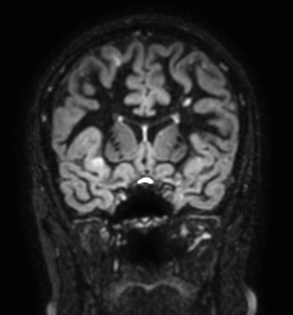
3D T2w FLAIR (reformat)
![3D T2w FLAIR (reformat)]()
-
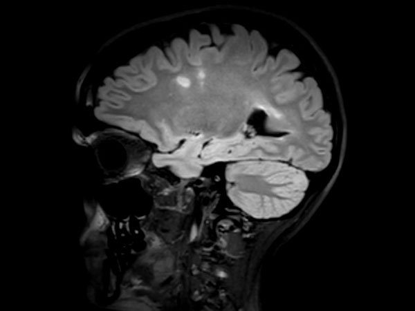
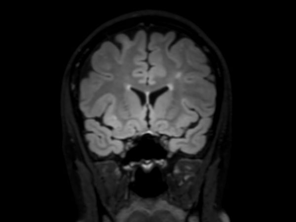
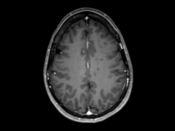
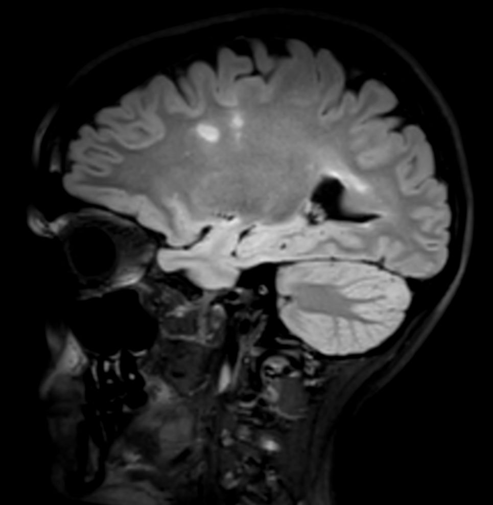
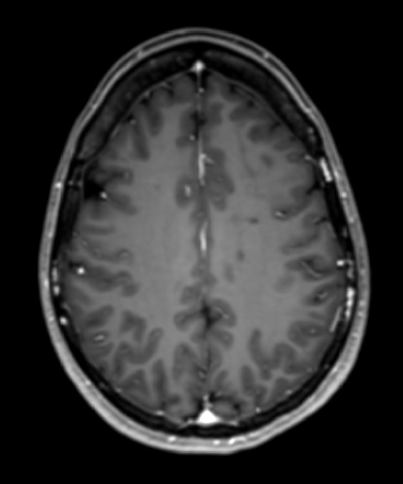
3D T2w FLAIR
![3D T2w FLAIR]()
-
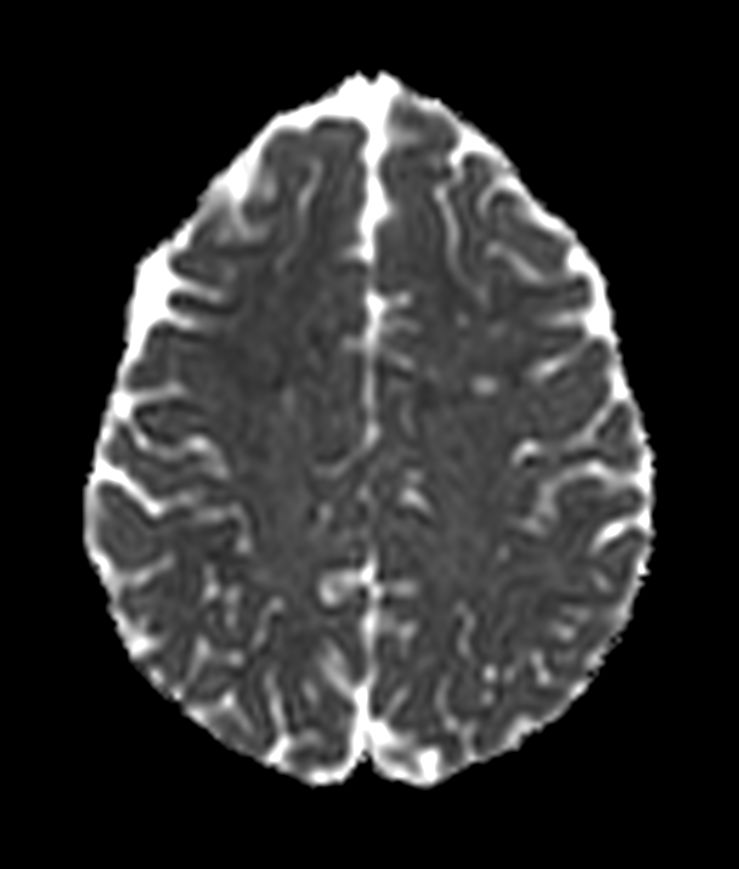
3D Double IR (reformat)
![3D Double IR (reformat)]()
-
3D Double IR (reformat)
![3D Double IR (reformat)]()
-
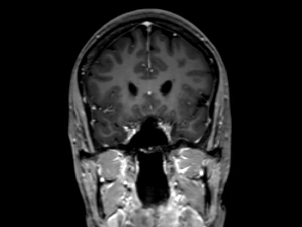
3D Double IR
![3D Double IR]()
-
3D T1w FFE +Gado (reformat)
![3D T1w FFE +Gado (reformat)]()
-
3D T1w FFE +Gado (reformat)
![3D T1w FFE +Gado (reformat)]()
-
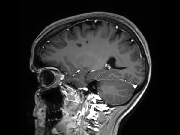
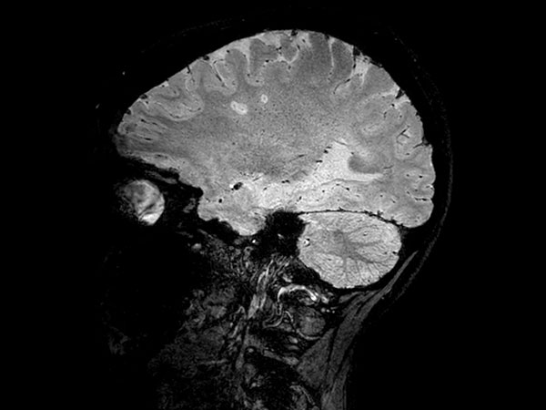
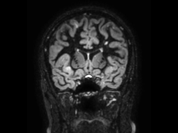
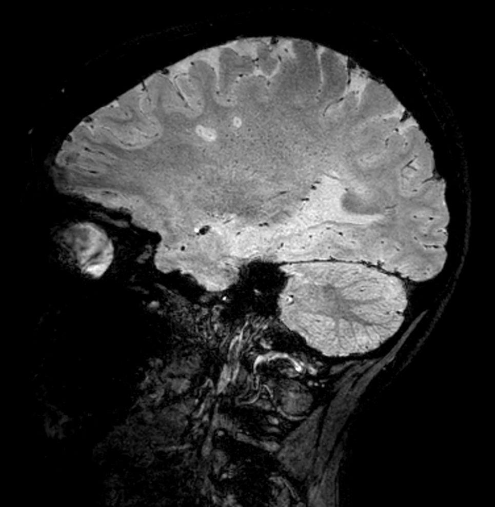
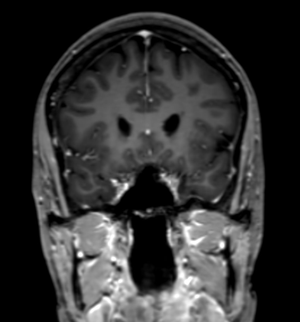
3D T1w FFE +Gado
![3D T1w FFE +Gado]()
-
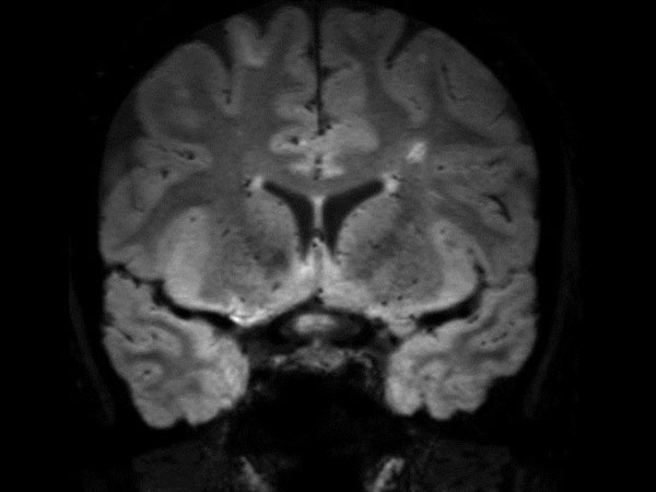
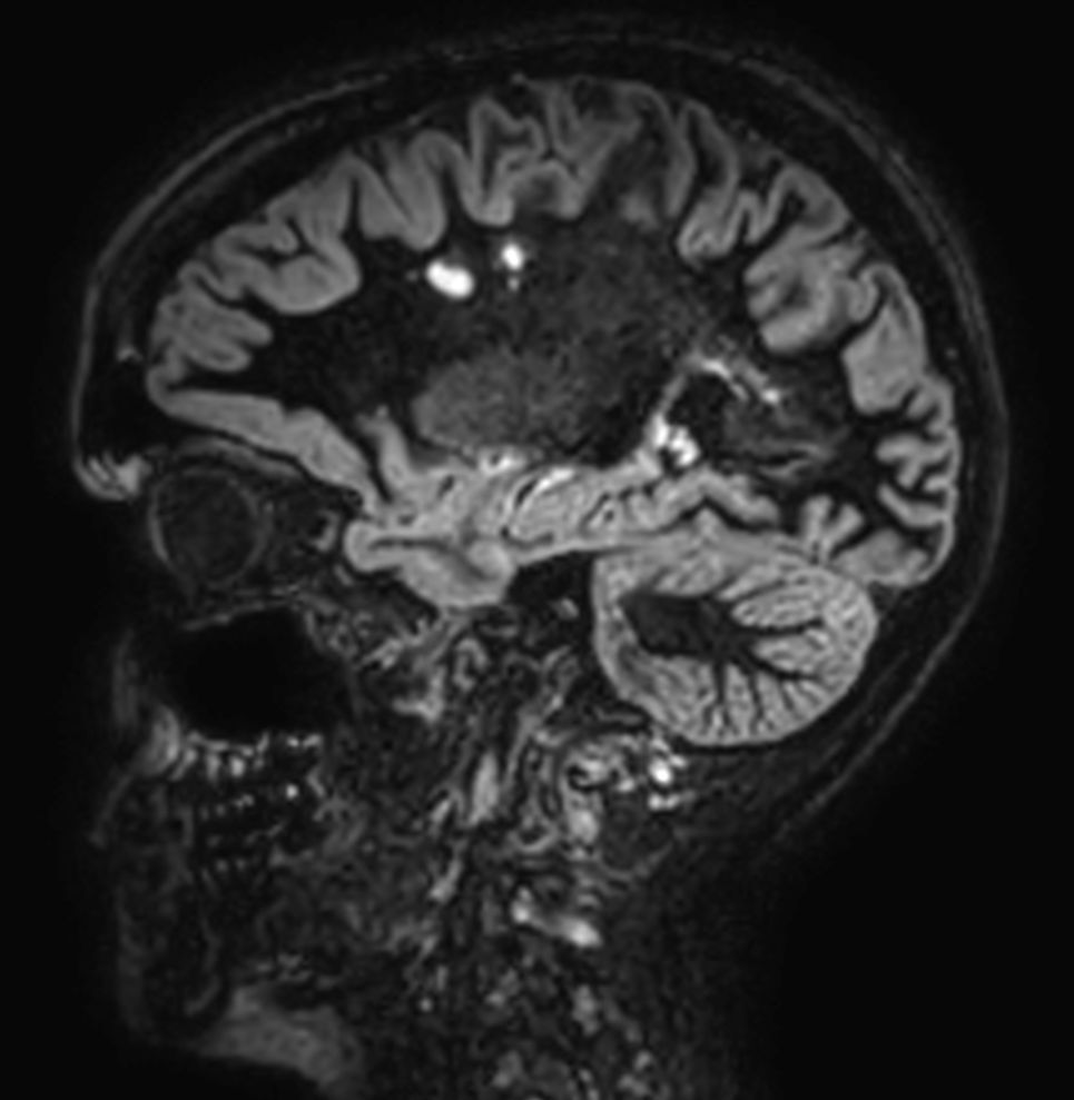
FLAIR*
![FLAIR*]()
Download ExamCards
FLAIR star protocol for Multiple SclerosisMore Information
- FieldStrength article: UBC researchers advance their multiple sclerosis imaging
- FieldStrength article: High quality imaging in MS, stroke and brain tumor
- FieldStrength article: Scan fast and have more time for advanced MRI techniques
- FieldStrength article: UVM brain MRI protocols upgraded with latest methods
- FieldStrength article: Imaging small cerebral aneurysms using non-invasive MR angiography
- FieldStrength article: Recently adopted methods for neuro MR improve efficiency and confidence
- FieldStrength article: Ingenia 3.0T delivers high performance MRI to the busy practice at DMG
- FieldStrength article: Fast MRI examinations and more comfort for patients
- FieldStrength article: MRI In-bore solutions
- Brochure: Clinical applications
- Video: Recording of webinar Dr. Nickerson on Neuro improvements
*Results from case studies are not predictive of results in other cases. Results in other cases may vary.