Go back
Comprehensive Liver exam of focal liver lesions
Patient information
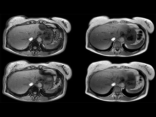
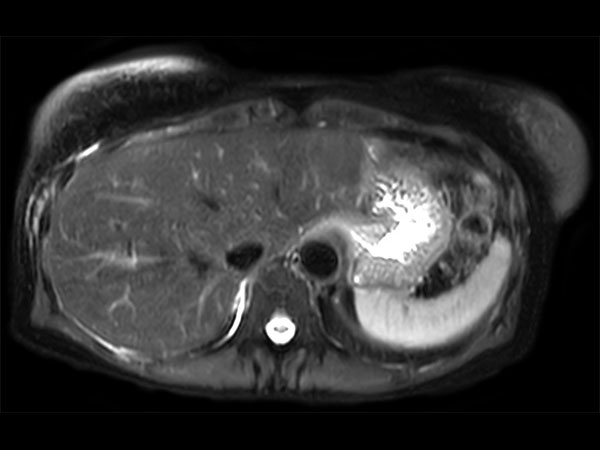
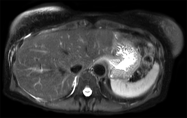
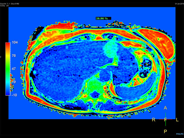
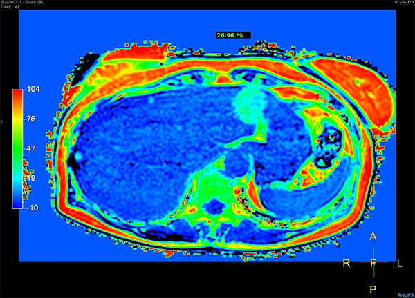
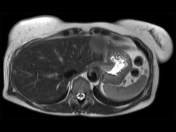
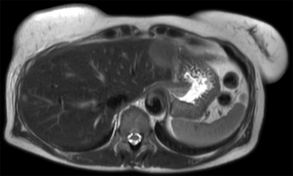
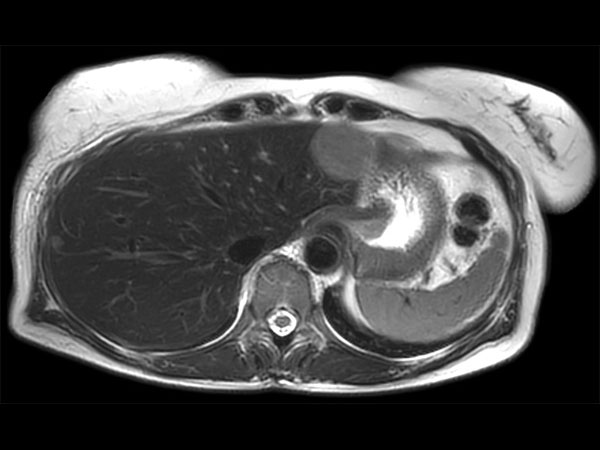
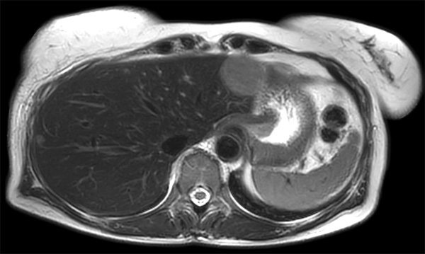
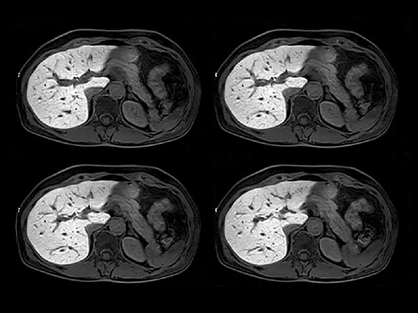
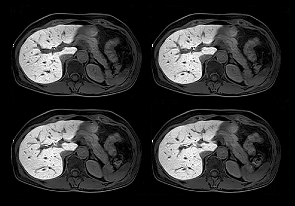
Patient with multiple liver lesions showing up hyperintense on T2-weighted images. The high spatial resolution of MultiVane XD can be appreciated by comparing the images demonstrating the two hyperintense focal liver lesions. mDIXON Quant was performed for quantification of intralesional and hepatic parenchymal fat content. The fat content in the liver parenchyma was normal. The lesion has a fat fraction of 25-30%. Histopathologic diagnosis was hepatocellular adenoma.Gallery

Download ExamCards
Free breathing abdominal imaging Comprehensive Liver imaging Advanced Liver imagingMore Information
- Brochure: Clinical applications
- FieldStrength article: How liver MR imaging gets boost from latest techniques
*Results from case studies are not predictive of results in other cases. Results in other cases may vary.