Go back
Multi-parametric MR of the prostate
Patient information
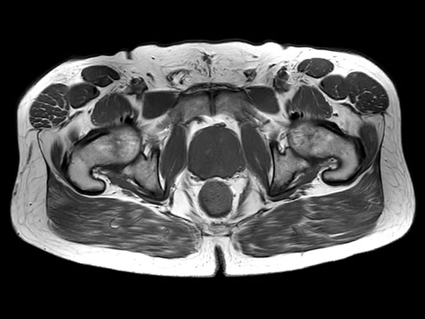
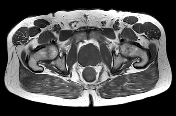
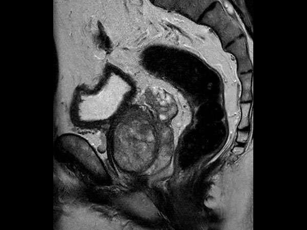
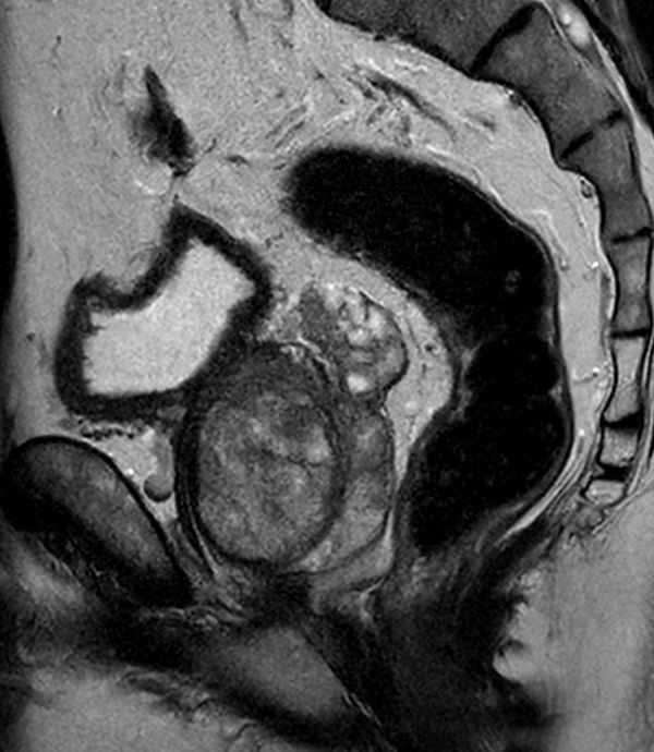
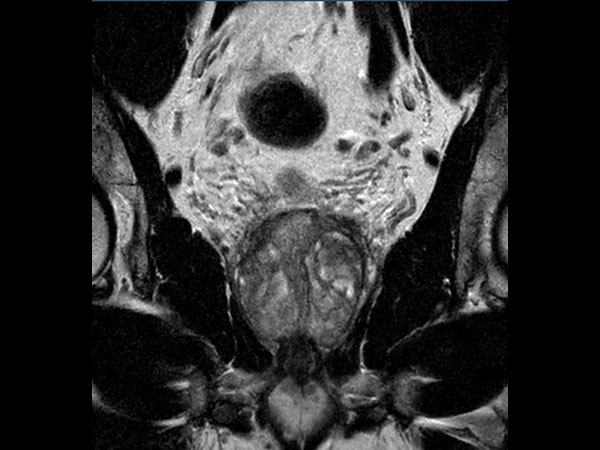
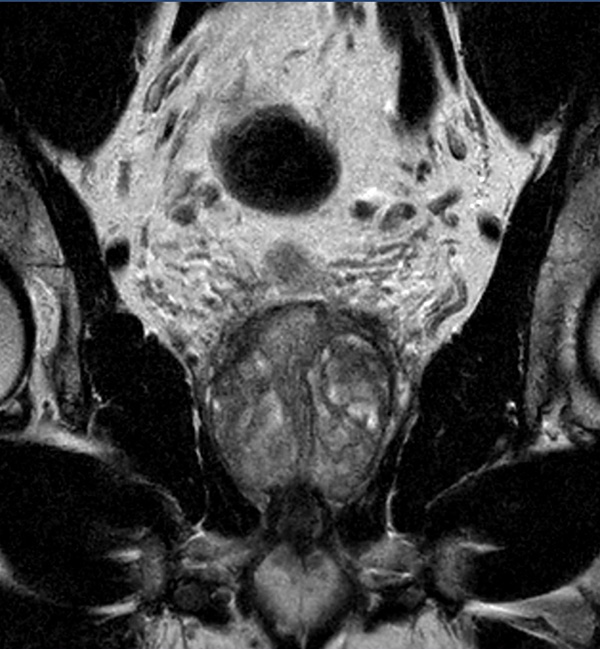
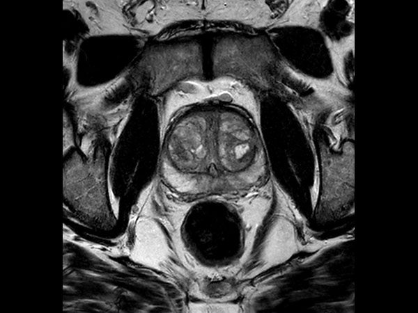
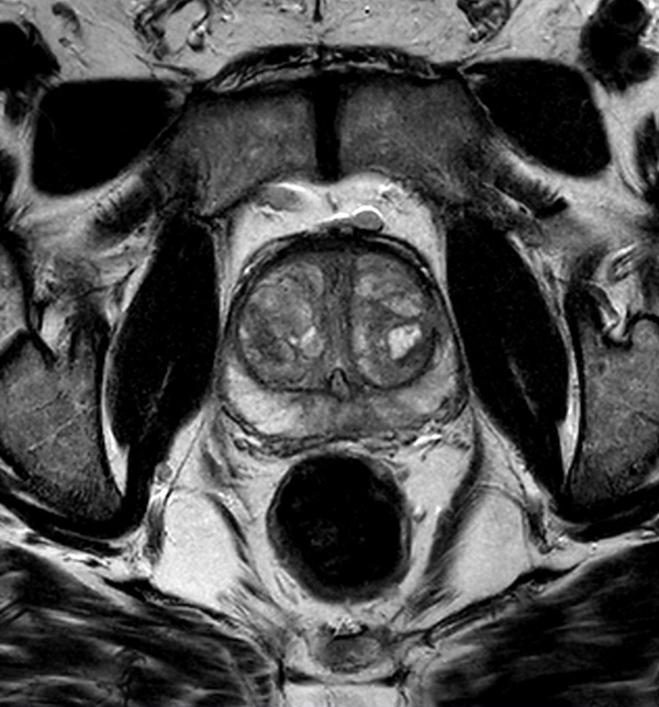
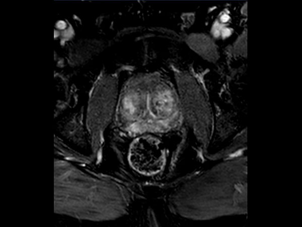
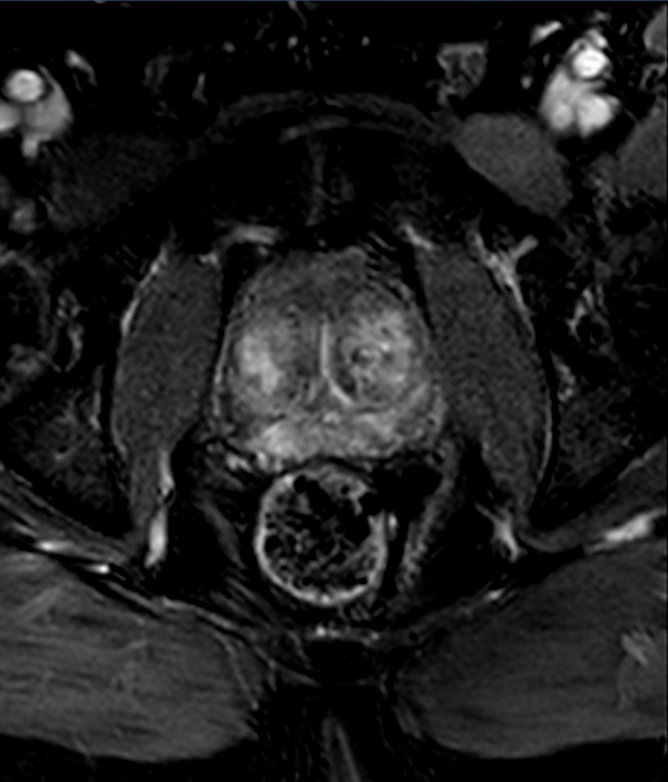
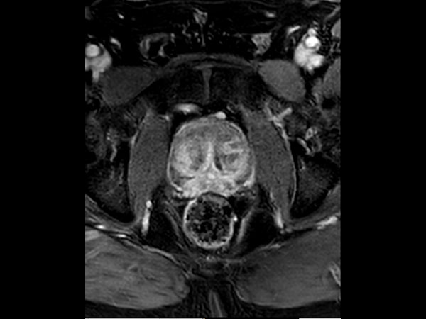
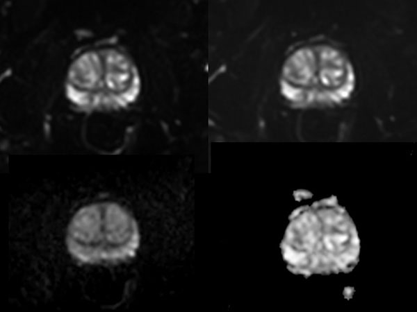
A 60-year-old male with elevated PSA and weak urinary stream underwent MRI. The exam includes high resolution DWI and ADC mapping as well as dynamic imaging. The prostate gland measures 5.2 x 4.4 x 5.9 cm in maximal transverse, AP, and craniocaudal dimensions, respectively, corresponding to an approximate glandular volume of 70 ml. Heterogeneous nodular hypertrophy is seen along the central transitional zone, with hypointense pseudo capsule, indicative of BPH, without dominant T2-hypointense nodules. Patchy T2-hypointense foci are noted throughout the peripheral zone bilaterally at the base, mid-gland and apex, with total PI-RADS score 6, so probably benign. No dominant nodular areas of restricted diffusion are evident. A geographic T2-hyperintense focus in the peripheral zone at the right base to mid-gland, paramidline shows asymmetric restricted diffusion, total PI-RADS score 10. No dominant lesions, greater than 1 cm. Clinical correlation and follow-up are advised.Gallery


More Information
- FieldStrength article: Ingenia 3.0T delivers high performance MRI to the busy practice at DMG
- FieldStrength article: The power of MRI and MRI/US biopsy to aid in prostate cancer diagnosis
- Compressed SENSE in practice: Doubling exam slots with no sacrifice to image quality
- White paper: mDIXON XD - The next generation fat free imaging
- Brochure: Clinical applications
*Results from case studies are not predictive of results in other cases. Results in other cases may vary.