Go back
dS Head 32ch coil - amyloid angiopathy
Patient information
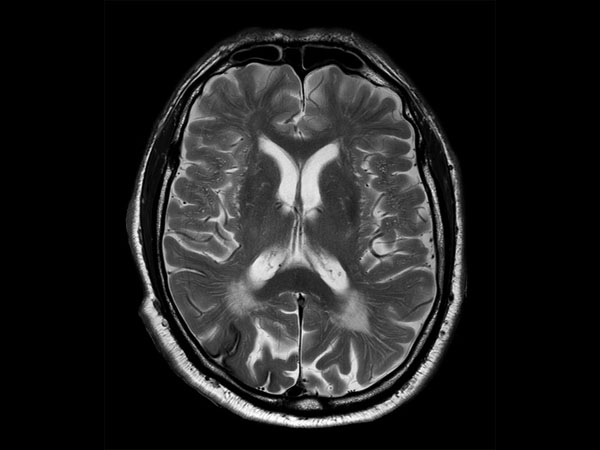
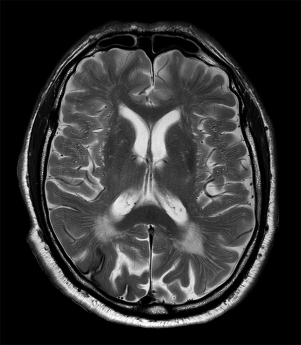
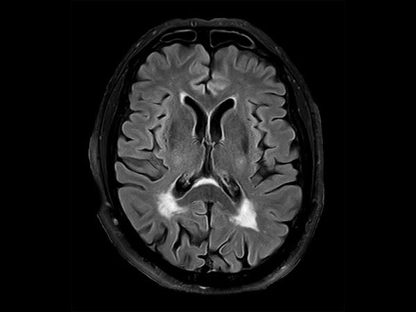
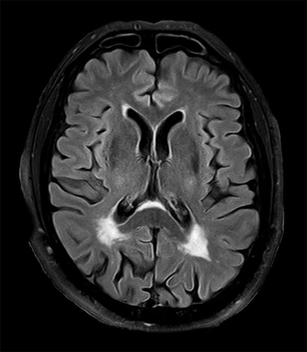
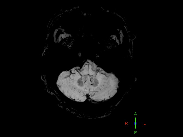
76-year-old male with history of amyloid angiopathy. Sagittal T1-weighted images shows chronic deep white matter ischemic changes. Axial T2-weighted and FLAIR demonstrate chronic ischemic changes and old hemorrhage / hemosiderin staining. Axial Venous BOLD (Susceptibility Weighted Imaging) image show chronic ischemic changes and old hemorrhage / hemosiderin staining. Imaging appears consistent with amyloid angiopathy, no other intracranial lesions are found.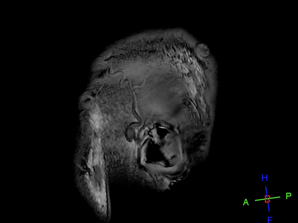
More Information
- Brochure: Clinical applications
- Video: MRI Neuro Highlights and Clinical Advancements at Dent Neurologic Institute
- FieldStrength article: Ingenia 3.0T delivers high performance MRI to the busy practice at DMG
- FieldStrength article: Recently adopted methods for neuro MR improve efficiency and confidence
- FieldStrength article: High quality imaging in MS, stroke and brain tumor
- FieldStrength article: Imaging small cerebral aneurysms using non-invasive MR angiography
- FieldStrength article: UVM brain MRI protocols upgraded with latest methods
*Results from case studies are not predictive of results in other cases. Results in other cases may vary.